Up Block11 Prev Next

 上一張相片  下一張相片  索引頁
block11_15.jpg - Fig. 6 & 7, NK-2-c, k-2-a, Pituitary Gland, anterior lobe, HEThis photomicrograph shows a region of the anterior lobe. The  acidophils are readily identified by the acidophilic staining of  their cytoplasm, in contrast to the basophils whose  cytoplasm is clearly basophilic. Chromophobes are also  very numerous in this field. The cytoplasm stains poorly in  contrast to that of the acidophils and basophils. The cells  are arranged in cords and clumps, between which are  capillaries. Some of the capillaries can be recognized, but  most are in a collapsed state and difficult to visualize at this  magnification.

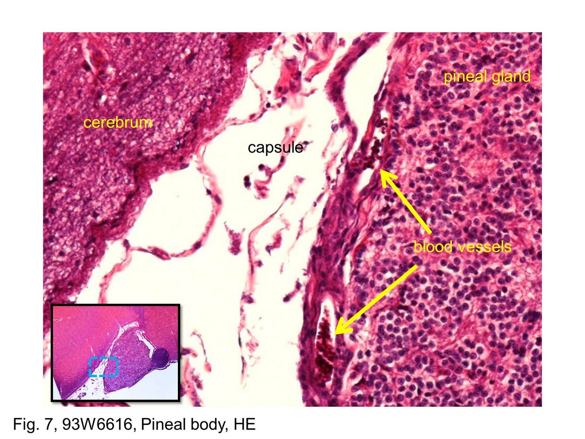
Fig. 7, 93W6616, Pineal body, HE This slide shows the pineal gland is surrounded by a very thin capsule that is formed by the pia mater. Connective tissue extends from the capsule into the substance of the gland. Blood vessels, generally small arteries and veins, course through the connective tissue. Adjacent to the pineal gland is the cerebrum.This slide shows the pineal gland is surrounded by a very thin capsule that is formed by the pia mater. Connective tissue extends from the capsule into the substance of the gland. Blood vessels, generally small arteries and veins, course through the connective tissue. Adjacent to the pineal gland is the cerebrum.
block11_07block11_09block11_11block11_12block11_14block11_15block11_17block11_18block11_20block11_22block11_30

支援訊息