|
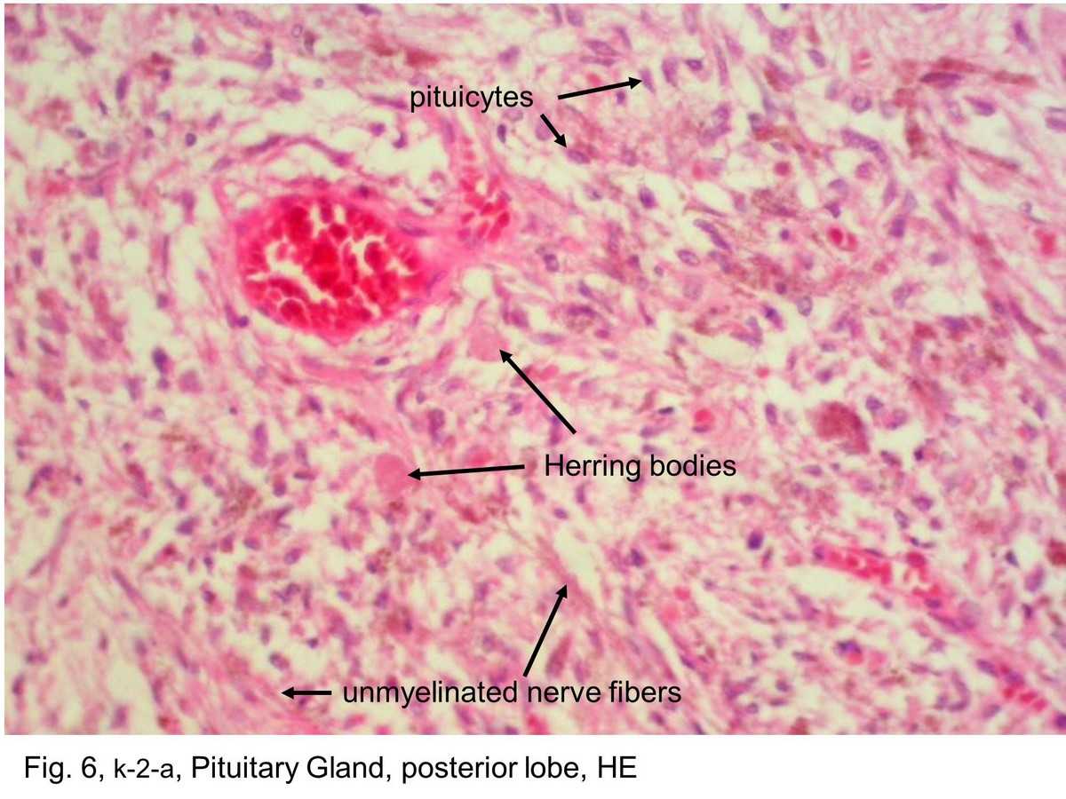 |
| Fig. 6, k-2-a, Pituitary Gland, posterior lobe, HE The posterior lobe seen here contains the nuclei inside the cells called pituicytes, and unmyelinated nerve fibers extended from the nuclei of the hypothalamus. The pituicytes are comparable with neuroglial cells of the central nervous system. The nuclei are round to oval. In H&E preparations such as this, the cytoplasm of the pituicyte cannot be distinguished from the unmyelinated nerve fibers. The hormones of the posterior lobe are formed in the hypothalamic soma and pass via the nerve fibers to the posterior lobe, where they are stored in the expanded nerve terminal portion of the nerve fibers. The stored neurosecretory material appears as Herring bodies. In H&E preparations, the Herring bodies simply appear as small islands of eosin-stained substance. | |||||||||||
支援訊息