Up Block11 Prev Next

 上一張相片  下一張相片  索引頁
block11_07.jpg - Fig. 2, NK-1-a, parathyroid gland, HE.Two parenchymal cell types can be distinguished in routine  H&E sections: chief cells (principal cells) and oxyphil cells.  The chief cells are more numerous. They contain a spherical  nucleus surrounded by a small amount of cytoplasm.  Oxyphil cells are less numerous. They are conspicuously  larger than chief cells but have a slightly smaller and more  intensely staining nucleus. Their cytoplasm stains with eosin,  and the boundaries between the cells are usually well  marked.

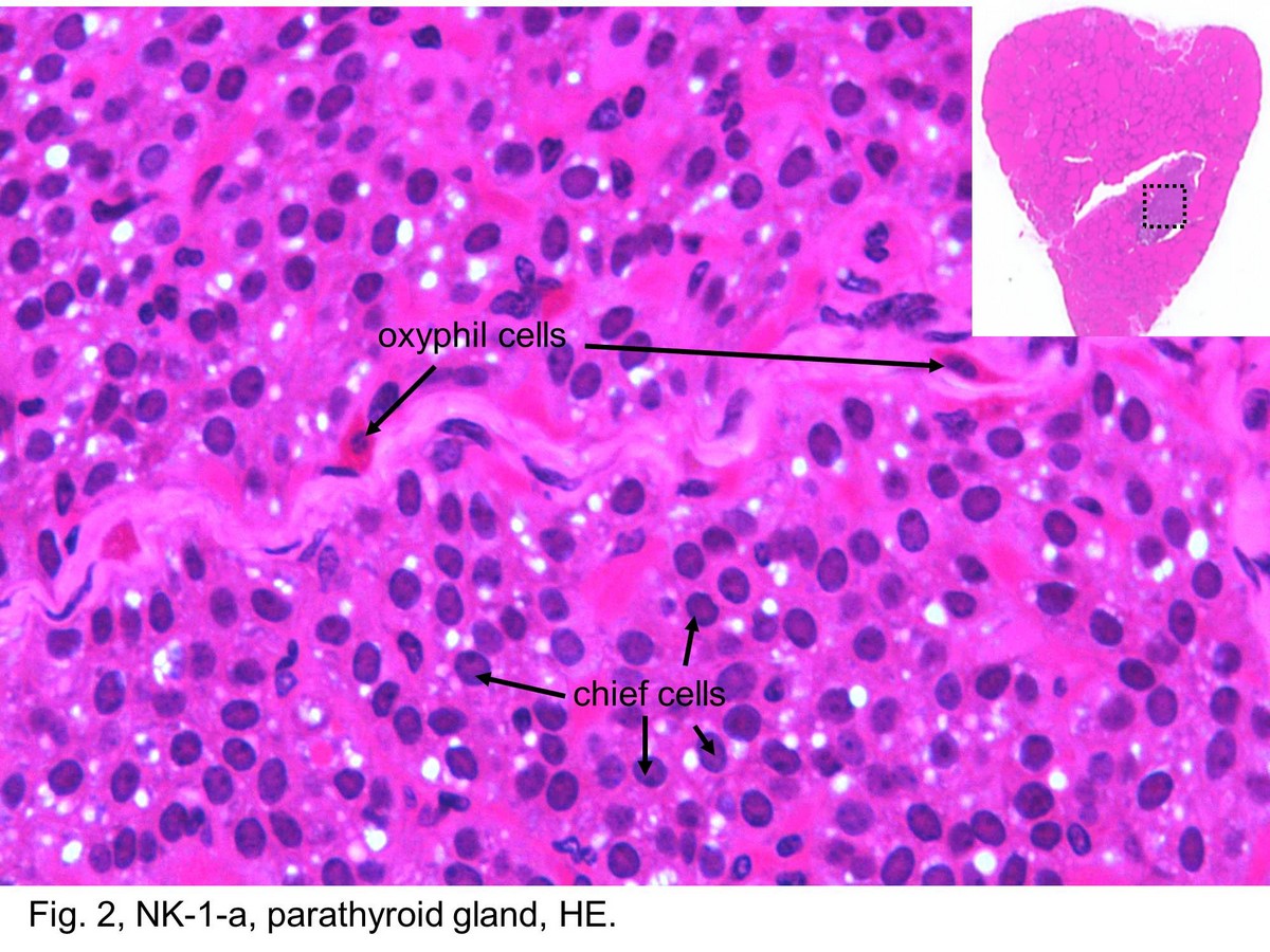
Fig. 2, NK-1-a, parathyroid gland, HE. Two parenchymal cell types can be distinguished in routine H&E sections: chief cells (principal cells) and oxyphil cells. The chief cells are more numerous. They contain a spherical nucleus surrounded by a small amount of cytoplasm. Oxyphil cells are less numerous. They are conspicuously larger than chief cells but have a slightly smaller and more intensely staining nucleus. Their cytoplasm stains with eosin, and the boundaries between the cells are usually well marked.
block11_01block11_02block11_03block11_04block11_05block11_07block11_09block11_11block11_12block11_14block11_15

支援訊息