Up Block11 Prev Next

 上一張相片  下一張相片  索引頁
block11_22.jpg - Fig. 11, K-3-a, Pineal gland (Epiphysis cerebri), Monkey, HE  This micrograph shows at higher magnification of  parenchyma of the pineal gland. Within the gland at the light  microscopic level there are two specific cell types. One cell  type represents the pinealocytes are by far the most  numerous. Pinealocytes are modified neurons. Their nuclei  are spherical and are relatively lightly stained because of the  amount of euchromatin that they contain. The second cell  type is the interstitial cell or glial cell that constitutes a  relatively small percentage of the cells in the gland. Their  nuclei are smaller and more elongate than those of the  pinealocytes.

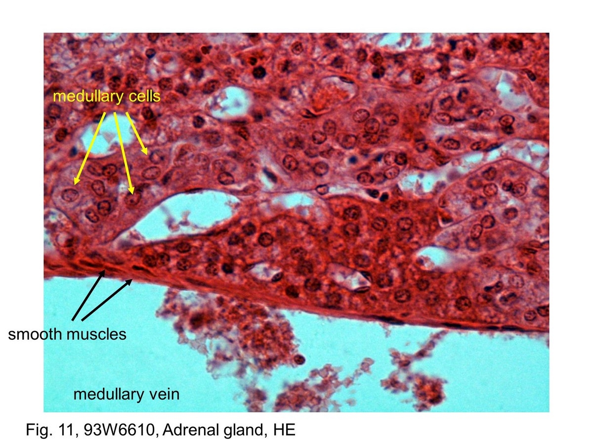
Fig. 11, 93W6610, Adrenal gland, HE This micrograph shows a medullary vein that drains the adrenal medulla. The smooth muscle of the tunica media is readily seen here as being arranged in bundles and appears in cross section. There is no discrete tunica adventitia in the smaller medullary veins. Instead, its connective tissue blends in with surrounding structures. Ganglion cells could be found in proximity to the wall of the medullary vein. They are large cells with a moderately basophilic cytoplasm. The cytoplasm of the medullary cells may stain with different intensity. The cytoplasm of some cells is very poorly stained, appearing almost clear, whereas others show greater intensity of eosin staining.
block11_14block11_15block11_17block11_18block11_20block11_22block11_30 

支援訊息