Up Block11 Prev Next

 上一張相片  下一張相片  索引頁
block11_17.jpg - Fig. 8 & 9, NK-2-c, k-2-a Pituitary Gland, posterior lobe, HEThe posterior lobe seen here contains the nuclei inside the cells  called pituicytes, and unmyelinated nerve fibers extended  from the nuclei of the hypothalamus. The pituicytes are  comparable with neuroglial cells of the central nervous system.  The nuclei are round to oval. In H&E preparations such as this,  the cytoplasm of the pituicyte cannot be distinguished from the  unmyelinated nerve fibers. The hormones of the posterior lobe  are formed in the hypothalamic soma and pass via the nerve  fibers to the posterior lobe, where they are stored in the  expanded nerve terminal portion of the nerve fibers. The  stored neurosecretory material appears as Herring bodies. In  H&E preparations, the Herring bodies simply appear as small  islands of eosin-stained substance.

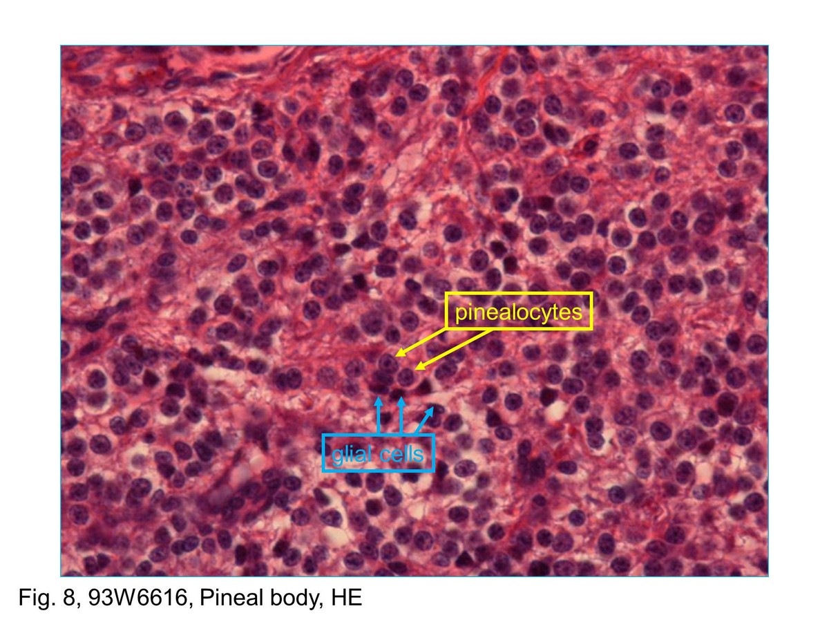
Fig. 8, 93W6616, Pineal body, HE This micrograph shows at higher magnification of parenchyma of the pineal gland. Within the gland at the light microscopic level there are two specific cell types. One cell type represents the pinealocytes are by far the most numerous. Pinealocytes are modified neurons. Their nuclei are spherical and are relatively lightly stained because of the amount of euchromatin that they contain. The second cell type is the interstitial cell or glial cell that constitutes a relatively small percentage of the cells in the gland. Their nuclei are smaller and more elongate than those of the pinealocytes.
block11_09block11_11block11_12block11_14block11_15block11_17block11_18block11_20block11_22block11_30

支援訊息