Up Block11 Prev Next

 上一張相片  下一張相片  索引頁
block11_18.jpg - Fig. 8 & 9, NK-2-c, k-2-a Pituitary Gland, posterior lobe, HEThe posterior lobe seen here contains the nuclei inside the cells  called pituicytes, and unmyelinated nerve fibers extended  from the nuclei of the hypothalamus. The pituicytes are  comparable with neuroglial cells of the central nervous system.  The nuclei are round to oval. In H&E preparations such as this,  the cytoplasm of the pituicyte cannot be distinguished from the  unmyelinated nerve fibers. The hormones of the posterior lobe  are formed in the hypothalamic soma and pass via the nerve  fibers to the posterior lobe, where they are stored in the  expanded nerve terminal portion of the nerve fibers. The  stored neurosecretory material appears as Herring bodies. In  H&E preparations, the Herring bodies simply appear as small  islands of eosin-stained substance.

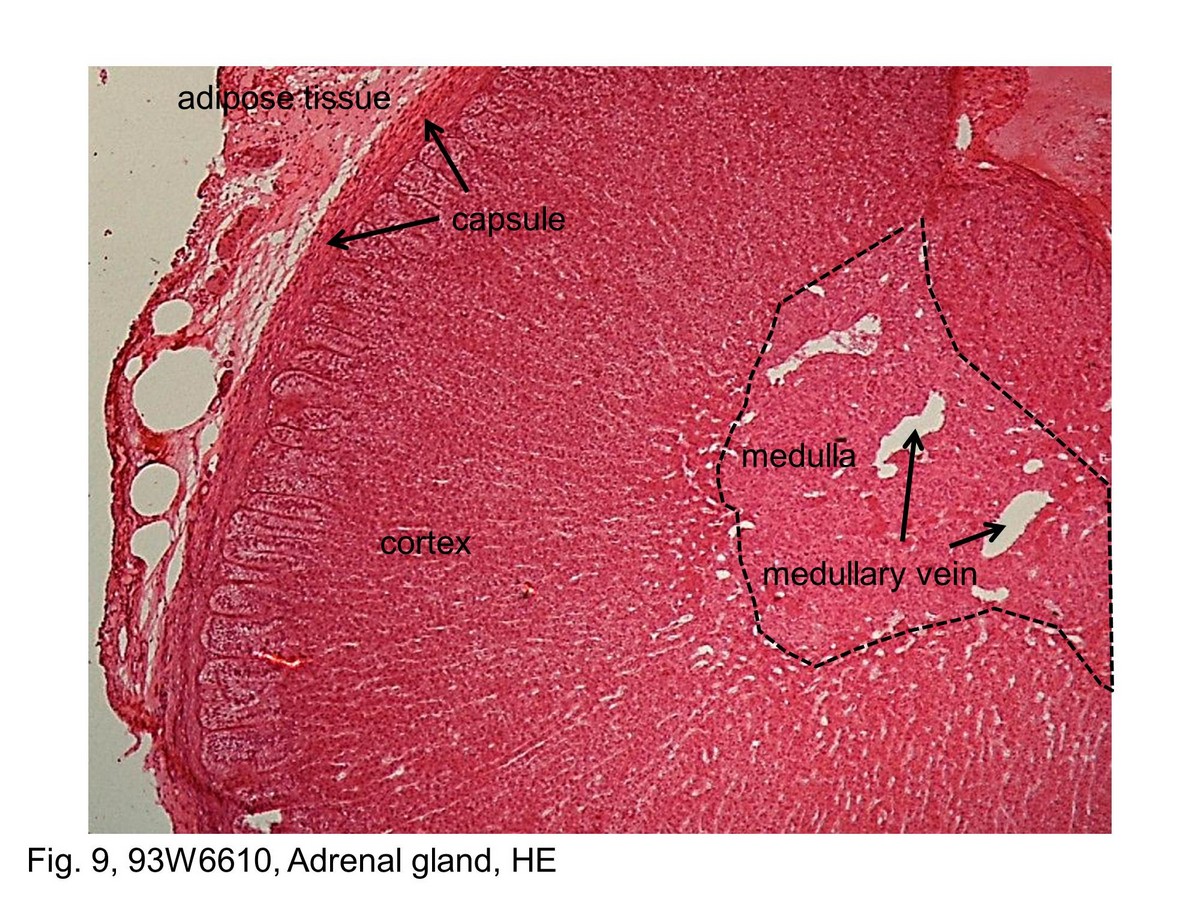
Fig. 9, 93W6610, Adrenal gland, HE This low-magnification micrograph of a section through the adrenal gland shows the outer capsule, which consists of dense connective tissue, the cortex and the underlying medulla. The cortex has a distinctly different appearance in both structural organization and staining characteristics of the medulla. A small amount of adipose tissue surrounds the capsule is seen at the lower portion of the micrograph. The corticomedullary boundary (dashed lines) has a wave-like contour. Within the medulla are a number of relatively large blood vessels. These are the medullary veins that drain both the cortex and the medulla.
block11_11block11_12block11_14block11_15block11_17block11_18block11_20block11_22block11_30

支援訊息