|
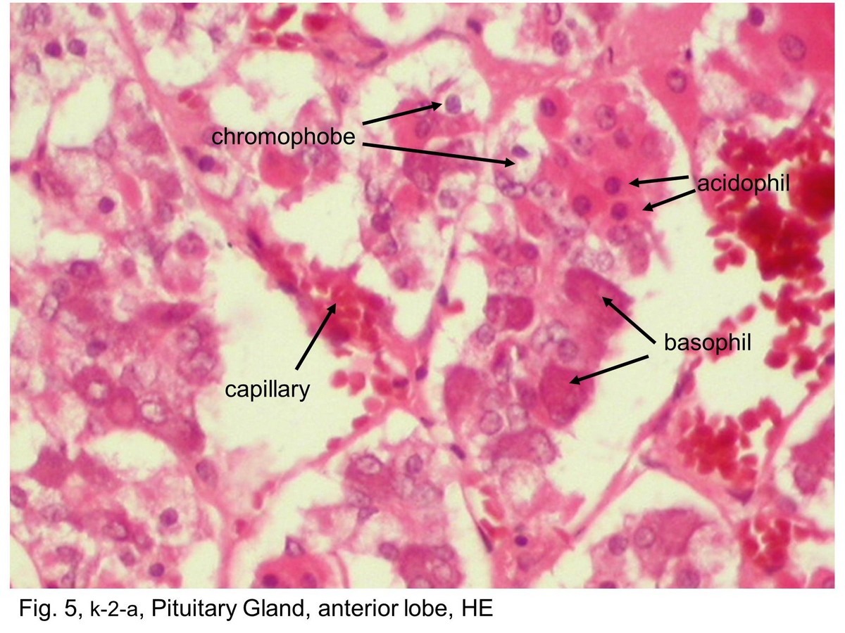 |
| Fig. 5, k-2-a, Pituitary Gland, anterior lobe, HE This photomicrograph shows a region of the anterior lobe. The acidophils are readily identified by the acidophilic staining of their cytoplasm, in contrast to the basophils whose cytoplasm is clearly basophilic. Chromophobes are also very numerous in this field. The cytoplasm stains poorly in contrast to that of the acidophils and basophils. The cells are arranged in cords and clumps, between which are capillaries. Some of the capillaries can be recognized, but most are in a collapsed state and difficult to visualize at this magnification. | |||||||||||
支援訊息