|
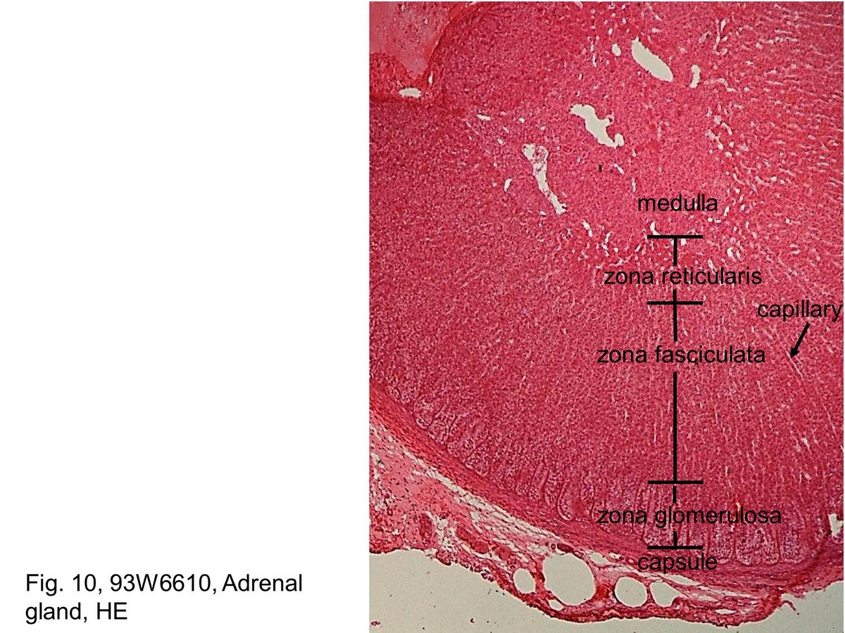 |
| Fig. 10, 93W6610, Adrenal gland, HE This is a higher magnification of a portion of the capsule and the full thickness of the cortex from an area in Figure 9. The zona glomerulosa is located at the outer part of the cortex, immediately under the capsule. The parenchyma of this zone consists of small cells that appear as oval groups of cells. The zona fasciculata consists of radially oriented cords and sheets of cells, usually two cells in width, that extend toward the medulla. Poor staining characteristic of cytoplasm of the zona fasciculata reflects more lipid droplets than those of the zona glomerulosa. Capillaries are located within the thin connective tissue and with the presence of red blood cells in their lumina. The cells of the zona reticularis are arranged in irregular anastomosing cords and contain only a small amount of lipid and, consequently, their cytoplasm stains with eosin. | ||||||||
支援訊息