Up Block8 Prev Next

 上一張相片  下一張相片  索引頁
block8_19.jpg - Fig.11 93W7125, Ureter (cs) , HE. As shown in this low-power orientation micrograph, the wall of the ureter consistsof a mucosa (Muc), a muscularis (Mus), and an adventitia(Adv). Note that the ureters are located behind theperitoneum of the abdominal cavity in their course to thebladder. Thus, a serosa (Ser) may be found covering aportion of the circumference of the tube. Also, because ofcontraction of the smooth muscle of the muscularis, theluminal surface is characteristically folded, thus creating astar-shaped lumen.

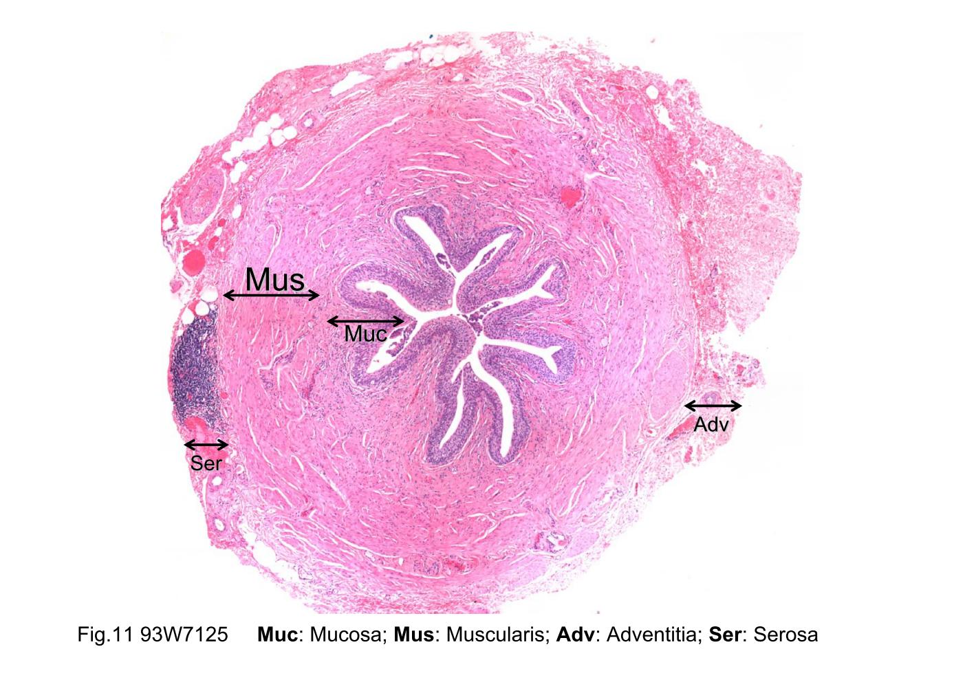
Fig.11 93W7125, Ureter (cs) , HE. As shown in this low- power orientation micrograph, the wall of the ureter consists of a mucosa (Muc), a muscularis (Mus), and an adventitia (Adv). Note that the ureters are located behind the peritoneum of the abdominal cavity in their course to the bladder. Thus, a serosa (Ser) may be found covering a portion of the circumference of the tube. Also, because of contraction of the smooth muscle of the muscularis, the luminal surface is characteristically folded, thus creating a star-shaped lumen.
block8_10block8_12block8_14block8_16block8_17block8_19block8_21block8_23block8_25block8_27block8_29

支援訊息