Up Block8 Prev Next

 上一張相片  下一張相片  索引頁
block8_14.jpg - Fig.7 The microscopic structure of renal medulla. Thisregion contains proximal and distal thick segments, thinsegments, and collecting tubules. All of the tubules areparallel, and all are cut in cross section; thus, they presentcircular profiles. Unfortunately, the tissue is shrunk seriouslyto be difficultly identified the different parts of tubule exceptthe collecting tubules (CT). The cells forming the collectingtubules are cuboidal and the boundaries between the cellsare usually evident (*); this serves as one of the mostdependable features for the identification of collecting tubules.

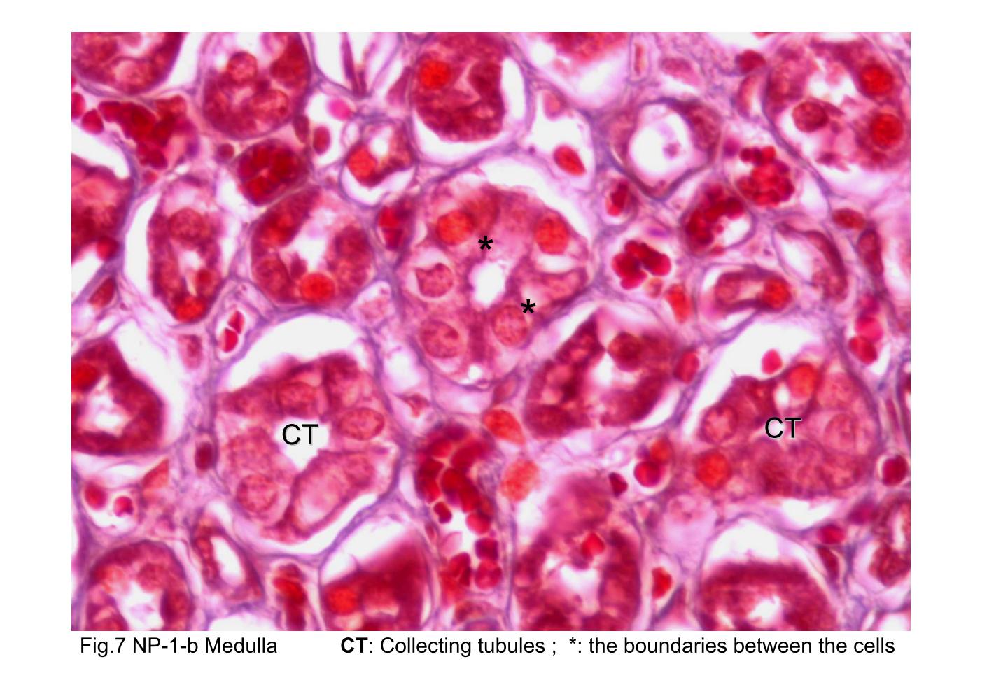
Fig.7 The microscopic structure of renal medulla. This region contains proximal and distal thick segments, thin segments, and collecting tubules. All of the tubules are parallel, and all are cut in cross section; thus, they present circular profiles. Unfortunately, the tissue is shrunk seriously to be difficultly identified the different parts of tubule except the collecting tubules (CT). The cells forming the collecting tubules are cuboidal and the boundaries between the cells are usually evident (*); this serves as one of the most dependable features for the identification of collecting tubules.
block8_04block8_06block8_08block8_10block8_12block8_14block8_16block8_17block8_19block8_21block8_23

支援訊息