Up Block8 Prev Next

 上一張相片  下一張相片  索引頁
block8_10.jpg - Fig 5. The microscopic structure of renal corpuscle. Therenal corpuscle appears as a spherical structure whichperiphery is composed of a thin capsule that encloses a narrowclear-appearing space, the urinary space (U), and a capillarytuft or glomerulus that appears as a large cellular mass. Thecapsule of the renal corpuscle, known as the renal orBowman's capsule, actually has two parts; a parietal layer,which is marked (BC), and a visceral layer. The parietal layerconsists of simple squamous epithelial cells. The proximalconvoluted tubules (PC) have a slightly larger outside diameterthan the distal convoluted tubules (DC) have. The proximaltubules have a brush border, whereas the distal tubules have acleaner, sharper luminal surface. Typically, fewer nuclei appearin a cross section of a proximal convoluted tubule than in anequivalent segment of a distal convoluted tubule.

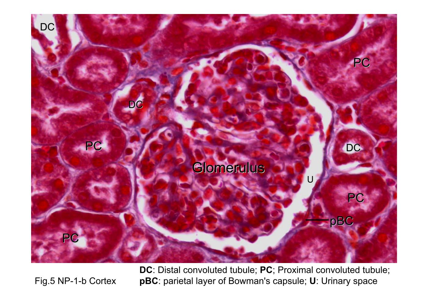
Fig 5. The microscopic structure of renal corpuscle. The renal corpuscle appears as a spherical structure which periphery is composed of a thin capsule that encloses a narrow clear-appearing space, the urinary space (U), and a capillary tuft or glomerulus that appears as a large cellular mass. The capsule of the renal corpuscle, known as the renal or Bowman's capsule, actually has two parts; a parietal layer, which is marked (BC), and a visceral layer. The parietal layer consists of simple squamous epithelial cells. The proximal convoluted tubules (PC) have a slightly larger outside diameter than the distal convoluted tubules (DC) have. The proximal tubules have a brush border, whereas the distal tubules have a cleaner, sharper luminal surface. Typically, fewer nuclei appear in a cross section of a proximal convoluted tubule than in an equivalent segment of a distal convoluted tubule.
block8_02block8_03block8_04block8_06block8_08block8_10block8_12block8_14block8_16block8_17block8_19

支援訊息