Up Block8 Prev Next

 上一張相片  下一張相片  索引頁
block8_25.jpg - Fig.14 93W7130, Urinary bladder human (cs), HE. Thetransitional epithelium (Ep) lining the bladder is seen on theleft. Beneath the epithelium is a relatively thick layer ofconnective tissue (CT) containing blood vessels of varioussizes. The epithelium and connective tissue constitute themucosa (Muc) of the bladder. The transitional epithelium isoften characterized by the presence of surface cells thatexhibit a "dome" shape. In addition, many of these surfacecells are binucleate (arrows). The muscularis (Mus) consistsof smooth muscle arranged as an inner longitudinal layer, amiddle circular layer, and an outer longitudinal layer, but it isless regularly arranged than the description indicates.

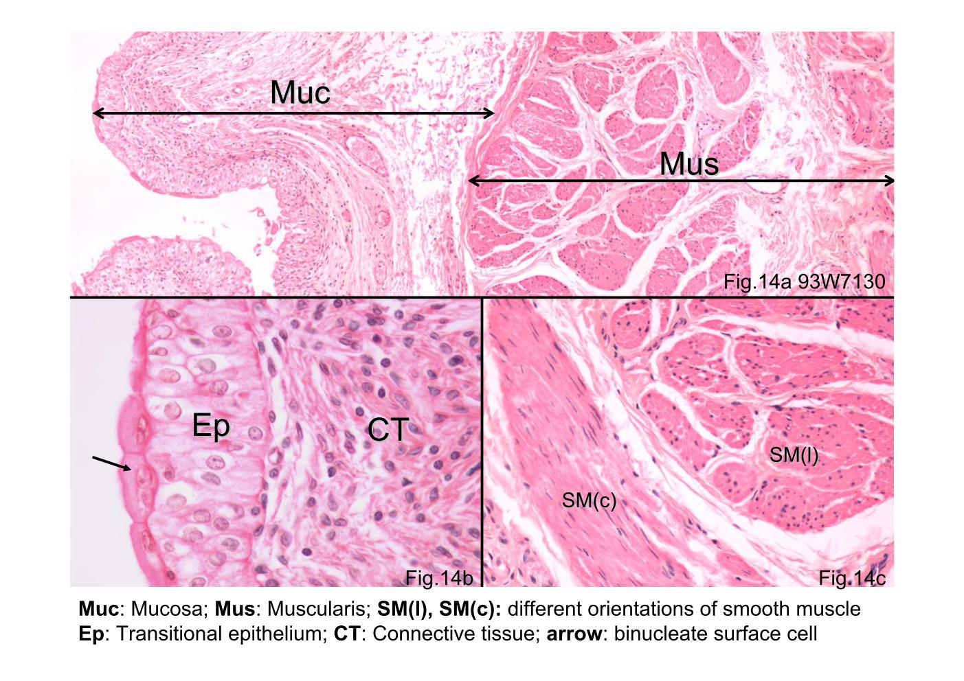
Fig.14 93W7130, Urinary bladder human (cs), HE. The transitional epithelium (Ep) lining the bladder is seen on the left. Beneath the epithelium is a relatively thick layer of connective tissue (CT) containing blood vessels of various sizes. The epithelium and connective tissue constitute the mucosa (Muc) of the bladder. The transitional epithelium is often characterized by the presence of surface cells that exhibit a "dome" shape. In addition, many of these surface cells are binucleate (arrows). The muscularis (Mus) consists of smooth muscle arranged as an inner longitudinal layer, a middle circular layer, and an outer longitudinal layer, but it is less regularly arranged than the description indicates.
block8_16block8_17block8_19block8_21block8_23block8_25block8_27block8_29block8_30block8_31 

支援訊息