Up Block8 Prev Next

 上一張相片  下一張相片  索引頁
block8_23.jpg - Fig.13 The photomicrograph of ureter mucosa. Thetransitional epithelium (Ep) and its supporting connectivetissue (CT) constitute the mucosa. The surface cells of thetransitional epithelium exhibit a rounded or dome-shapedprofile, and some are binucleate (arrow). The basal cells arethe smallest. The intermediate cells appear to consist ofseveral layers and are composed of cells larger in size thanthe basal cells but smaller than the surface cells.

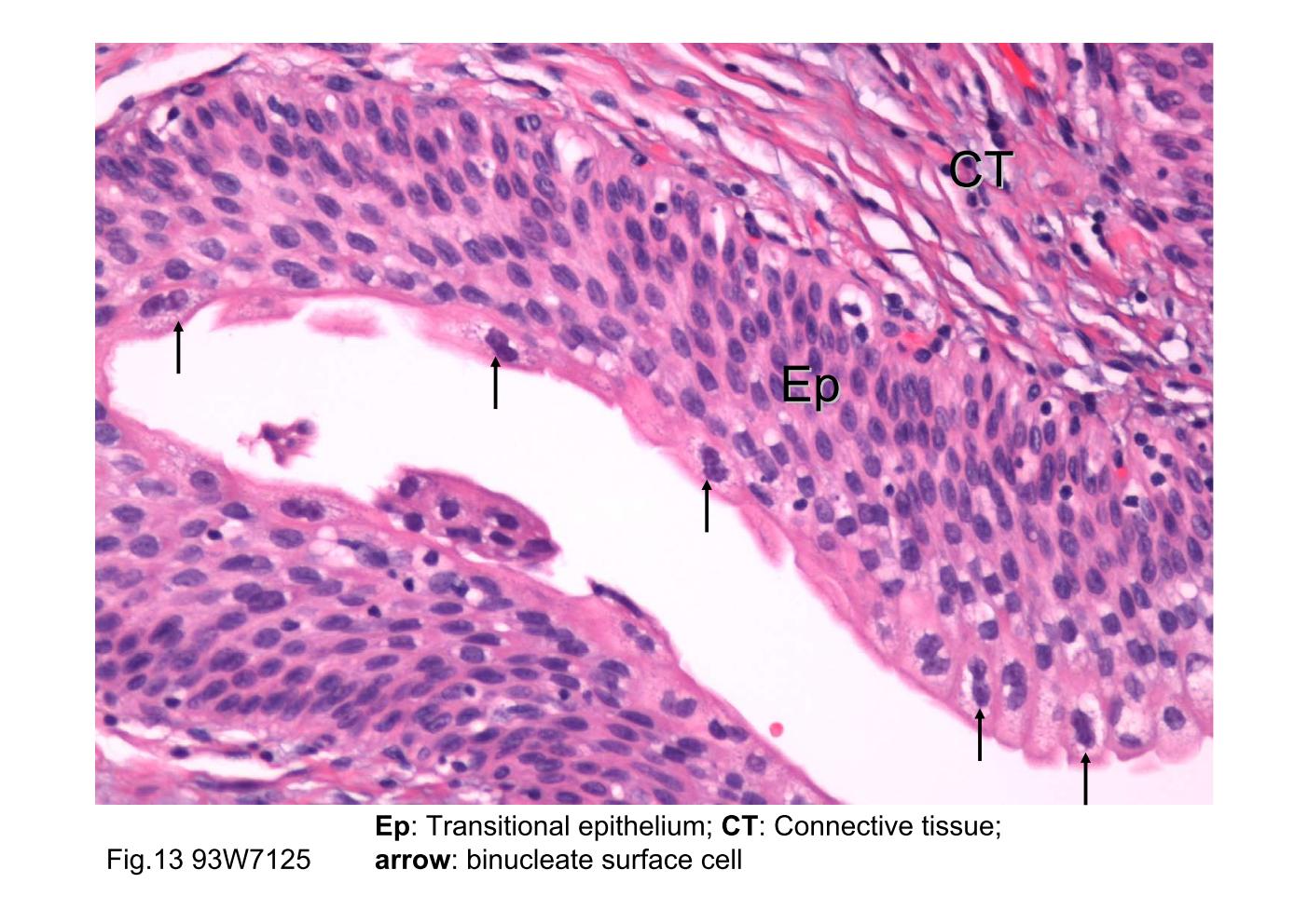
Fig.13 The photomicrograph of ureter mucosa. The transitional epithelium (Ep) and its supporting connective tissue (CT) constitute the mucosa. The surface cells of the transitional epithelium exhibit a rounded or dome-shaped profile, and some are binucleate (arrow). The basal cells are the smallest. The intermediate cells appear to consist of several layers and are composed of cells larger in size than the basal cells but smaller than the surface cells.
block8_14block8_16block8_17block8_19block8_21block8_23block8_25block8_27block8_29block8_30block8_31

支援訊息