Up Block8 Prev Next

 上一張相片  下一張相片  索引頁
block8_21.jpg - Fig.12 93W7125, Ureter (cs) , HE. The thick epitheliai liningis the transitional epithelium (Ep). The remainder of the wall ismade up of connective tissue (CT) and smooth muscle. Thetransitional epithelium and its supporting connective tissueconstitute the mucosa (Muc). A distinct submucosa is notpresent. The muscularis (Mus) is arranged as an innerlongitudinal layer (SM(l)), a middle circular layer (SM(c)), andan outer longitudinal layer (SM(l)). However, the outerlongitudinal layer is present only at the lower end of the ureter.By the way, there are blood vessels (BV) and lymphaticnodule (LN) in the serosa (Ser).

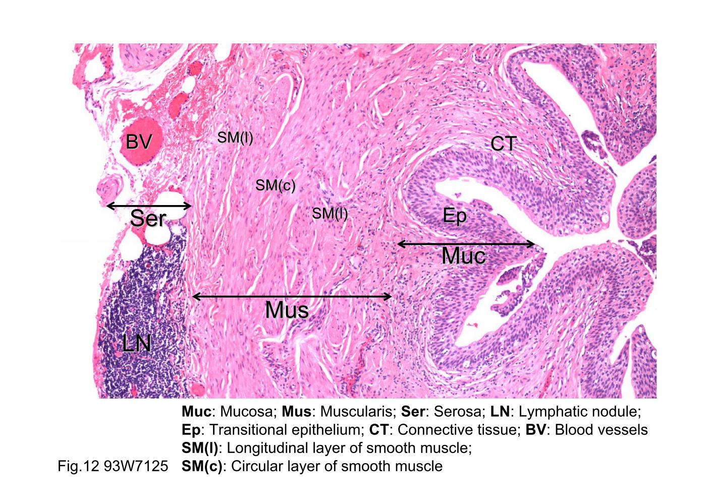
Fig.12 93W7125, Ureter (cs) , HE. The thick epitheliai lining is the transitional epithelium (Ep). The remainder of the wall is made up of connective tissue (CT) and smooth muscle. The transitional epithelium and its supporting connective tissue constitute the mucosa (Muc). A distinct submucosa is not present. The muscularis (Mus) is arranged as an inner longitudinal layer (SM(l)), a middle circular layer (SM(c)), and an outer longitudinal layer (SM(l)). However, the outer longitudinal layer is present only at the lower end of the ureter. By the way, there are blood vessels (BV) and lymphatic nodule (LN) in the serosa (Ser).
block8_12block8_14block8_16block8_17block8_19block8_21block8_23block8_25block8_27block8_29block8_30

支援訊息