|
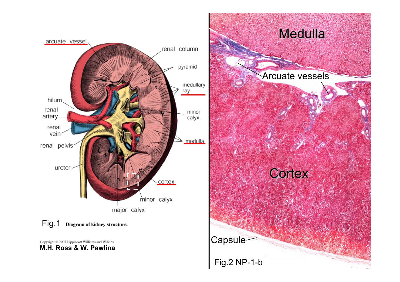 |
| Fig.1 Diagram of kidney structure. The diagram represents
a hemisection of a kidney, revealing its structural organization.
The white dashed line rectangle shows the orientation of
figure2. Fig.2 NP-1-b Kidney, human, PAS stain. PAS staining is mainly used for staining structures containing high proportion of carbohydrate macromolecules, typically found in connective tissues, mucus, and basal laminae to create a purple-magenta color. The lower part of the section is the cortex. It is easily distinguished from the upper portion above the white dashed line, the medulla. The arcuate vessels are located at the boundary between the cortex and the medulla. |
|||||||||||
支援訊息