Up Block8 Prev Next

 上一張相片  下一張相片  索引頁
block8_08.jpg - Fig 4. The microscopic structure of renal cortex. The renalcortex can be divided into regions referred to the corticallabyrinth (CL) and the medullary rays (MR). The corticallabyrinth contains the renal corpuscles (RC). Surrounding eachrenal corpuscle are the proximal and distal convoluted tubules,which are also part of the cortical labyrinth. The medullary raysare composed of groups of straight tubules oriented in thesame direction and appear to radiate from the medulla. Whenthe medullary rays are cut longitudinally, as they are in thisfigure, the tubules present elongated profiles. The medullaryrays contain proximal thick segments (descending limb ofHenle's loop), distal thick segments (ascending limbs of Henle'sloop), and collecting tubules. But they can¡¦t be identified well inthis slide.

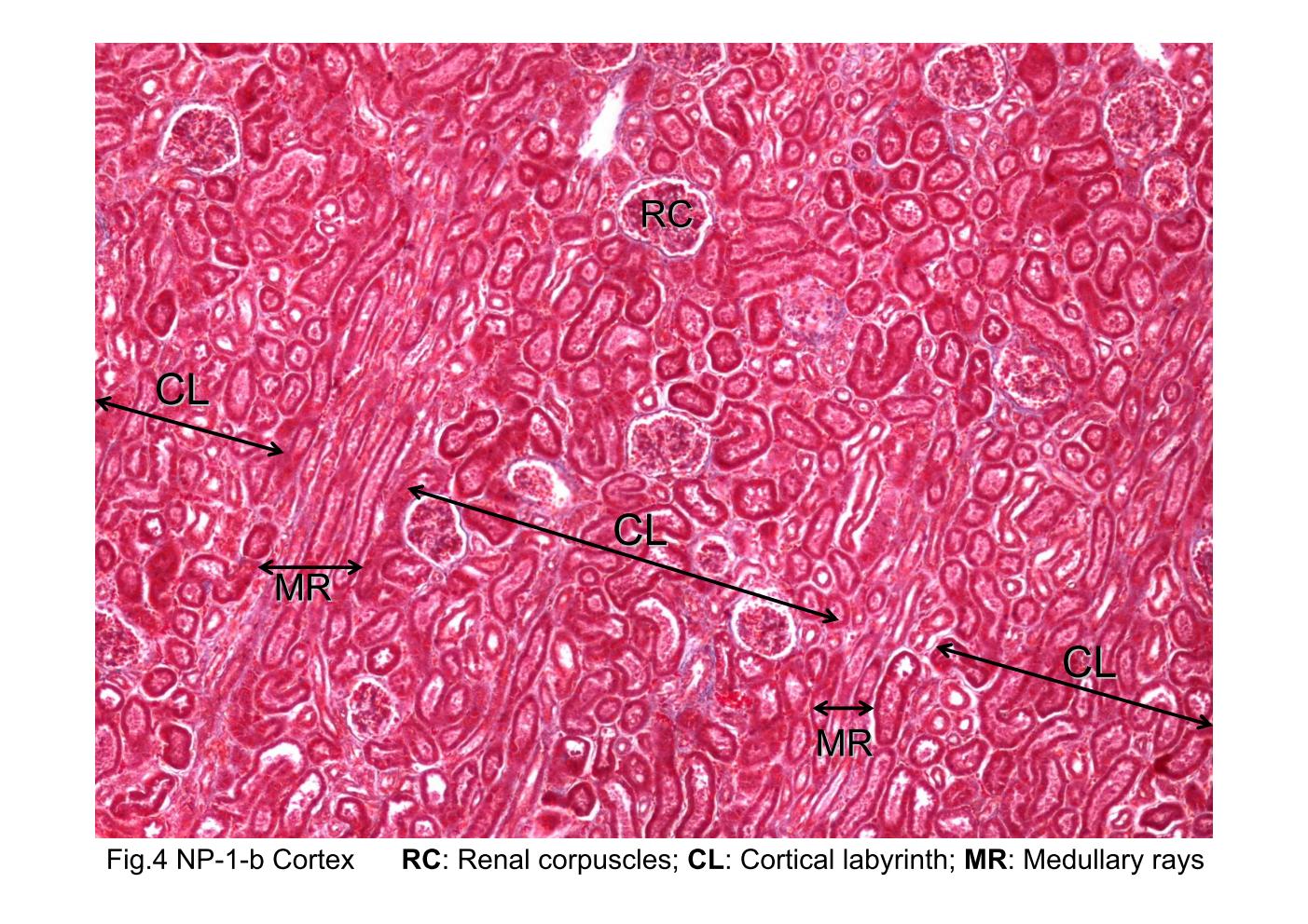
Fig 4. The microscopic structure of renal cortex. The renal cortex can be divided into regions referred to the cortical labyrinth (CL) and the medullary rays (MR). The cortical labyrinth contains the renal corpuscles (RC). Surrounding each renal corpuscle are the proximal and distal convoluted tubules, which are also part of the cortical labyrinth. The medullary rays are composed of groups of straight tubules oriented in the same direction and appear to radiate from the medulla. When the medullary rays are cut longitudinally, as they are in this figure, the tubules present elongated profiles. The medullary rays contain proximal thick segments (descending limb of Henle's loop), distal thick segments (ascending limbs of Henle's loop), and collecting tubules. But they can¡¦t be identified well in this slide.
block8_01block8_02block8_03block8_04block8_06block8_08block8_10block8_12block8_14block8_16block8_17

支援訊息