Up Block2 Prev Next

 上一張相片  下一張相片  索引頁
block3_16.jpg - Fig.8 93W3236 Reticular tissue m&nfr. The Manuel silvermethod makes the reticular fibers black, and the nuclear fastred stains the nuclei red. The main structural support for thelymph node is derived from the capsule and trabeculae, whichextend into the node. From these, a fine meshwork of reticularfibers extends throughout the node, providing a supportingframework for the mass of lymphocytes and accessory cellswithin the cortex and medullary cords. The reticular network isparticularly dense in the cortex, except for the follicular areaswhere it is relatively sparse. Compare this slide with the H&Estaining slide (93W6559) and find out the distribution ofreticular fibers only demonstrated by m&nfr staining.

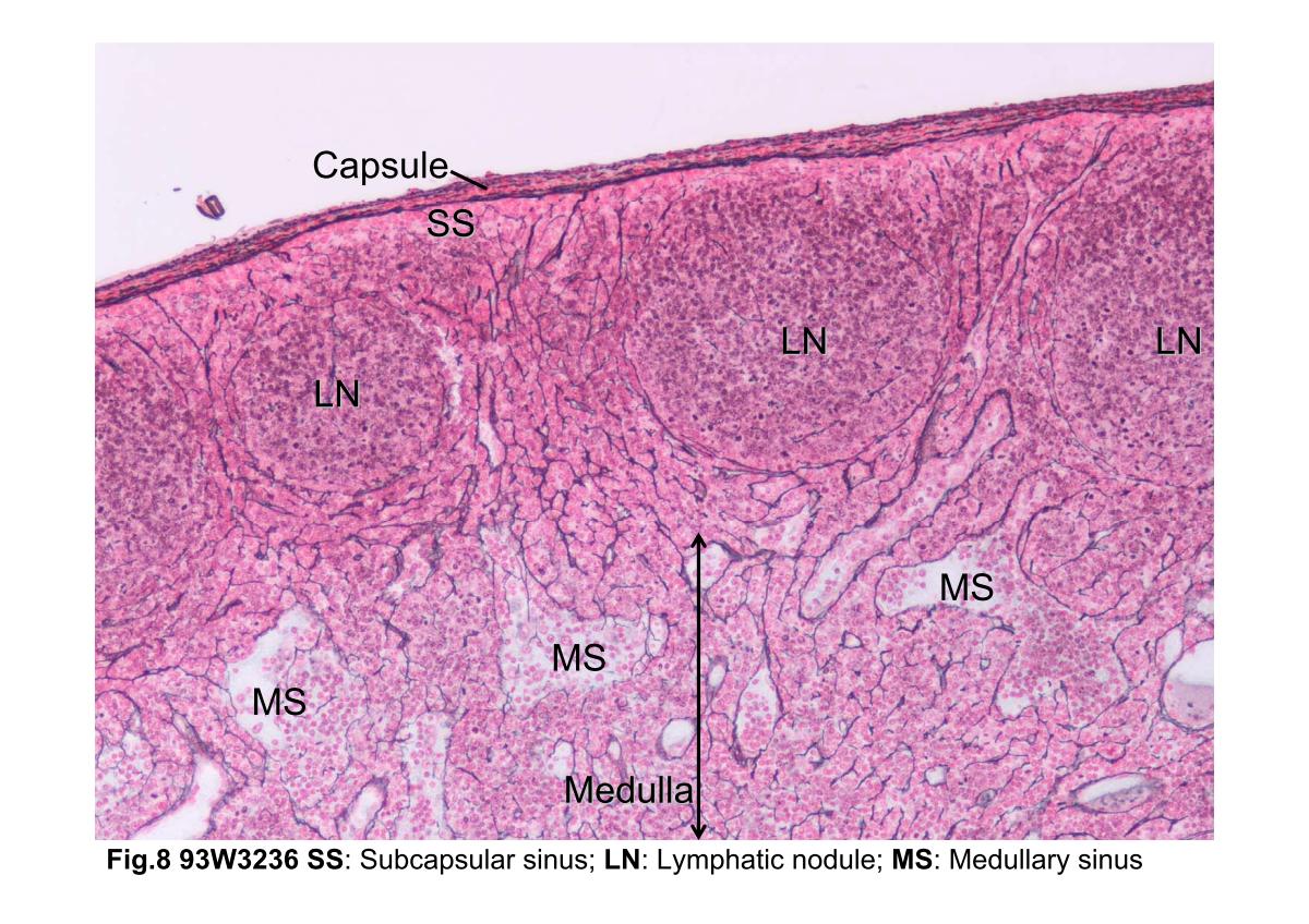
Fig.8 93W3236 Reticular tissue m&nfr. The Manuel silver method makes the reticular fibers black, and the nuclear fast red stains the nuclei red. The main structural support for the lymph node is derived from the capsule and trabeculae, which extend into the node. From these, a fine meshwork of reticular fibers extends throughout the node, providing a supporting framework for the mass of lymphocytes and accessory cells within the cortex and medullary cords. The reticular network is particularly dense in the cortex, except for the follicular areas where it is relatively sparse. Compare this slide with the H&E staining slide (93W6559) and find out the distribution of reticular fibers only demonstrated by m&nfr staining.
block3_06block3_08block3_10block3_12block3_14block3_16block3_18block3_20block3_22block3_24block3_26

支援訊息