|
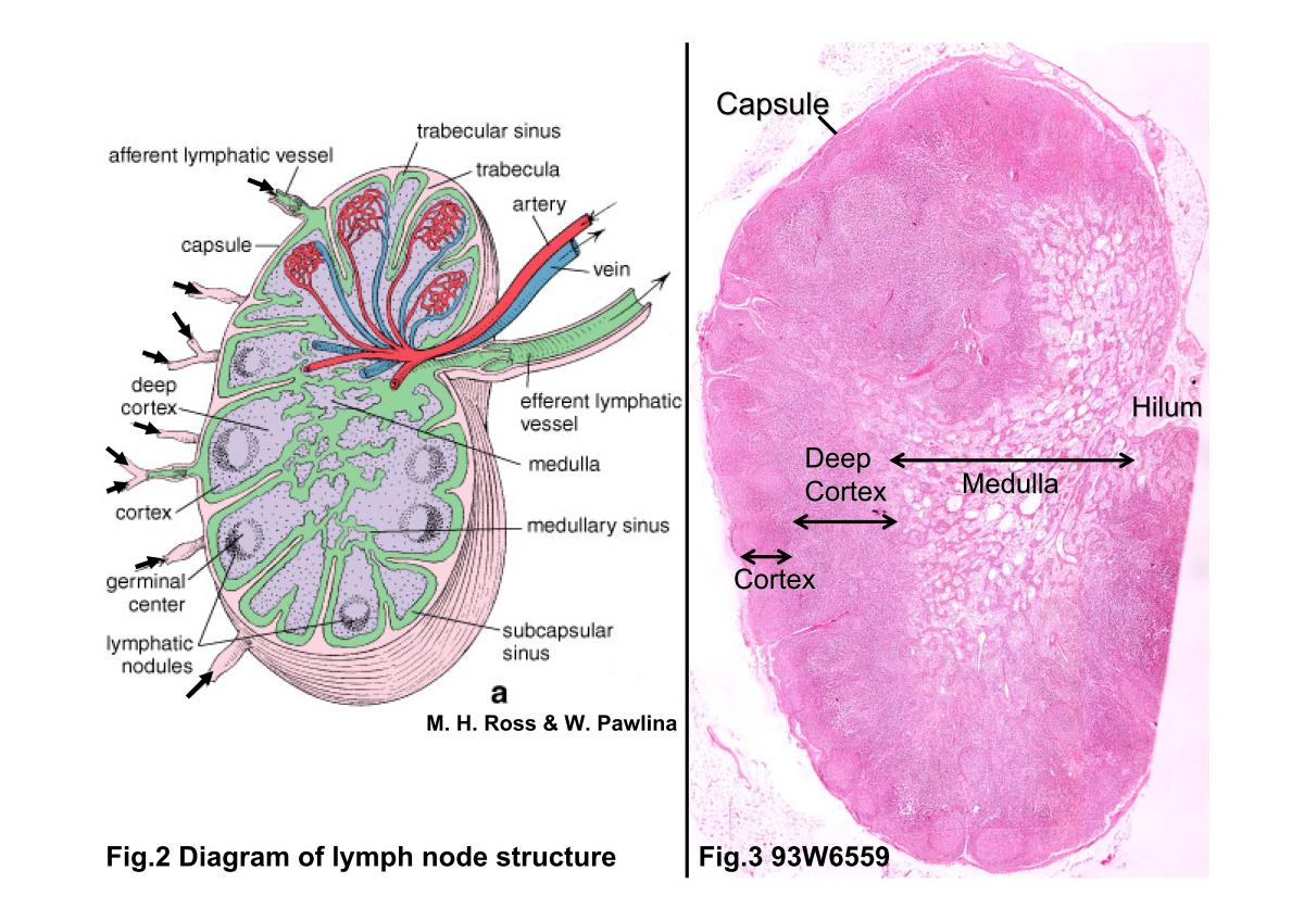 |
| Fig.2 Diagram of lymph node structure. Surrounding the
lymph node is a capsule of dense connective tissue from
which trabeculae (pink) extend into the substance of the node.
Under the capsule and adjacent to the trabeculae are,
respectively, the subcapsular sinus and the trabecular
lymphatic sinuses (green). Afferent lymphatic vessels (arrows)
penetrate the capsule and empty into the subcapsular
sinus. The subcapsular sinus and trabecular sinuses
communicate with the medullary sinuses. Fig.3 93W6559 Lymph node, H&E. The dense outer portion of the lymph node is the cortex. It consists of aggregations of lymphocytes organized as nodules and a nodule-free deep cortex. The innermost portion, the medulla, extends to the surface at the hilum, where blood vessels enter or leave and where efferent lymphatic vessels leave the node. Surrounding the lymph node is the capsule. |
|||||||||||
支援訊息