Up Block2 Prev Next

 上一張相片  下一張相片  索引頁
block3_08.jpg - Fig.4 Photomicrograph of a lymph node. This photo-micrograph shows the cortex, deep cortex, and medulla. Thecapsule is composed of dense connective tissue from whichtrabeculae (T) penetrate into the organ. The lymphatic nodules(LN) in the black dash line circles are the characteristic of theouter cortex. The deep cortex is nodule free. It consists ofdensely packed lymphocytes. In contrast to these areas, themedulla is a less dense area.

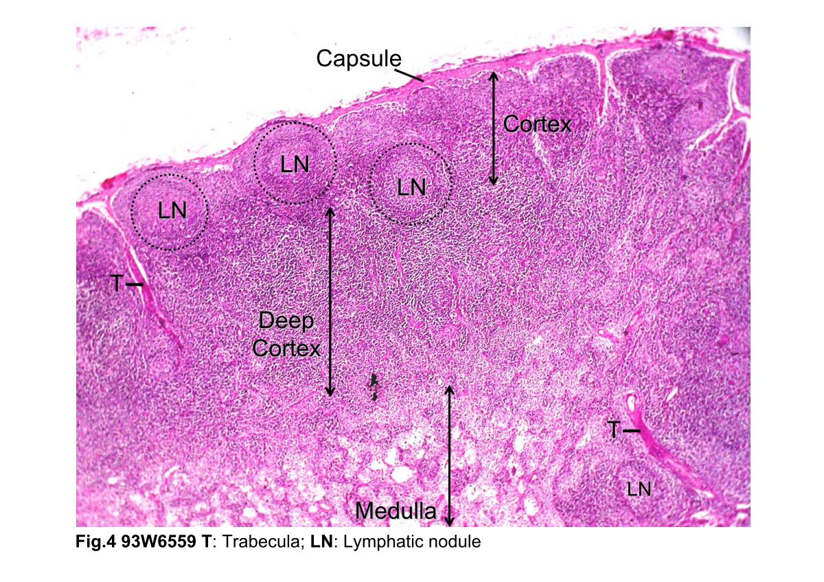
Fig.4 Photomicrograph of a lymph node. This photo- micrograph shows the cortex, deep cortex, and medulla. The capsule is composed of dense connective tissue from which trabeculae (T) penetrate into the organ. The lymphatic nodules (LN) in the black dash line circles are the characteristic of the outer cortex. The deep cortex is nodule free. It consists of densely packed lymphocytes. In contrast to these areas, the medulla is a less dense area.
block3_01block3_02block3_03block3_04block3_06block3_08block3_10block3_12block3_14block3_16block3_18

支援訊息