|
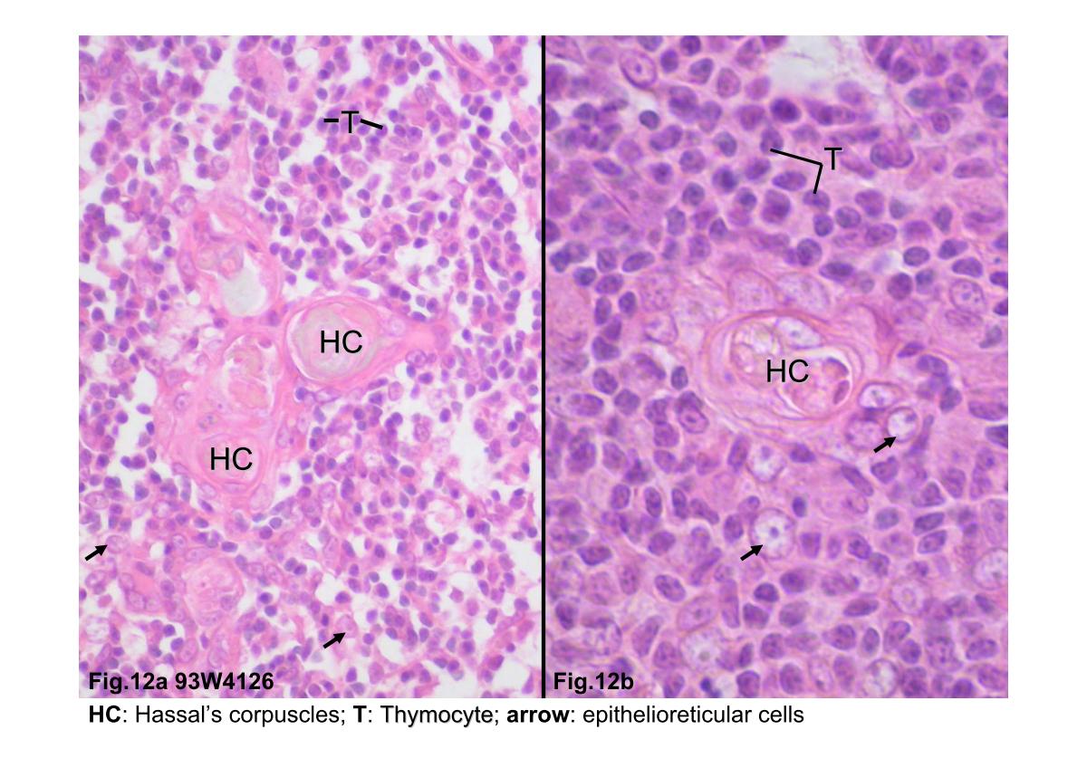 |
| Fig.12a Photomicrograph of thymus medulla. This high
magnification photomicrograph shows the medulla with
varying numbers of circular bodies called Hassall's corpuscles
(HC). The corpuscles are isolated masses of closely packed,
concentrically arranged epithelioreticular cells; these cells
exhibit flattened nuclei. In addition to numerous thymocytes
(T), the micrograph also shows epithelioreticular cells (arrows),
with their large, pale-staining nuclei. Fig.12b A higher magnification photomicrograph shows the basophilic thymocytes and the large, pale-staining epithelioreticular cells. |
|||||||||||
支援訊息