Up Block2 Prev Next

 上一張相片  下一張相片  索引頁
block3_20.jpg - Fig.10 93W6560 Spleen, H&E. This figure reveals at highermagnification of the spleen. The white pulp contains lymphatictissue that follows and ensheathes the central artery. The redpulp consists of splenic sinuses surrounded by splenic cords.The venous sinuses are filled with red blood cells and appearred in spleen, thus the name.

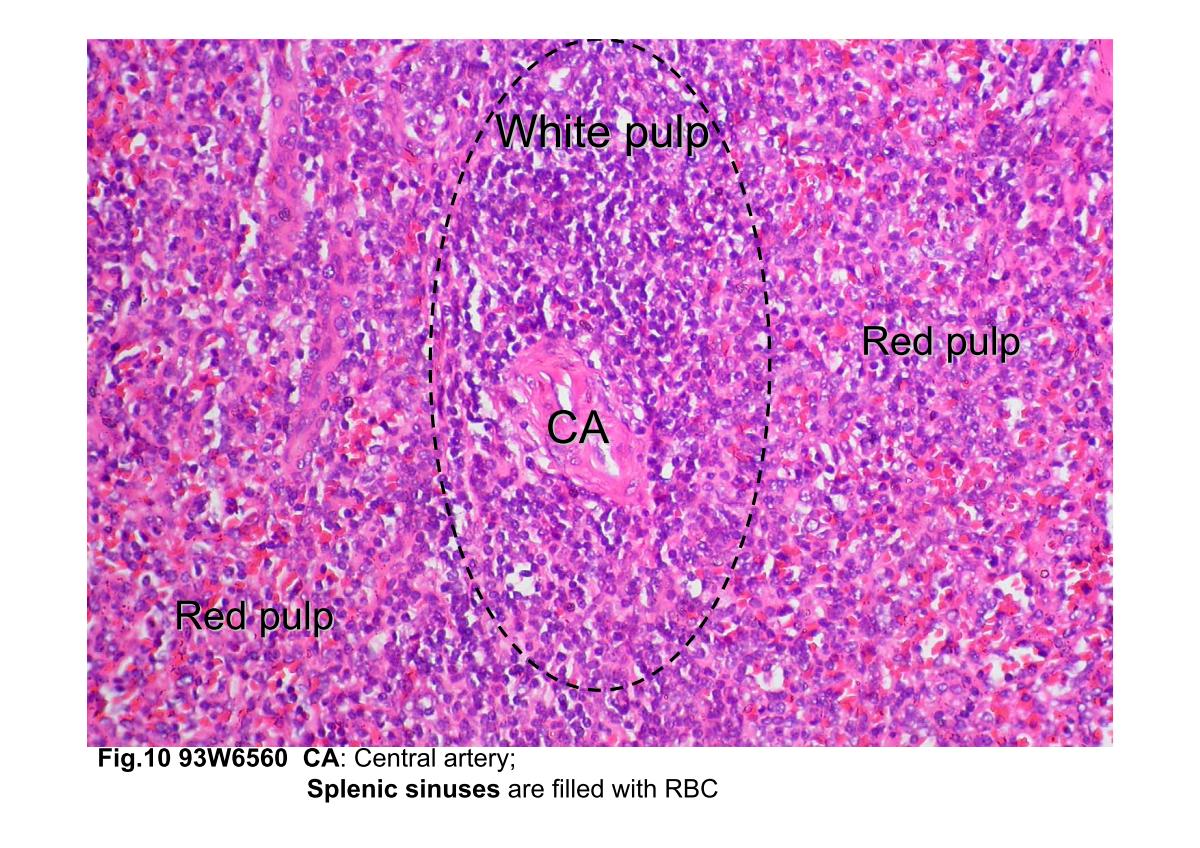
Fig.10 93W6560 Spleen, H&E. This figure reveals at higher magnification of the spleen. The white pulp contains lymphatic tissue that follows and ensheathes the central artery. The red pulp consists of splenic sinuses surrounded by splenic cords. The venous sinuses are filled with red blood cells and appear red in spleen, thus the name.
block3_10block3_12block3_14block3_16block3_18block3_20block3_22block3_24block3_26block3_28block3_29

支援訊息