Up Block2 Prev Next

 上一張相片  下一張相片  索引頁
block3_12.jpg - Fig.6 Photomicrograph of a lymphatic nodule. The capsuleis composed of dense connective tissue from whichtrabeculae (T) penetrate into the organ. Immediately below thecapsule is the subcapsular sinus (SS), which receives lymphfrom the afferent lymphatic vessels after they penetrate thecapsule. The subcapsular sinus is continuous with thetrabecular sinuses (TS) that course along the trabeculae (T).

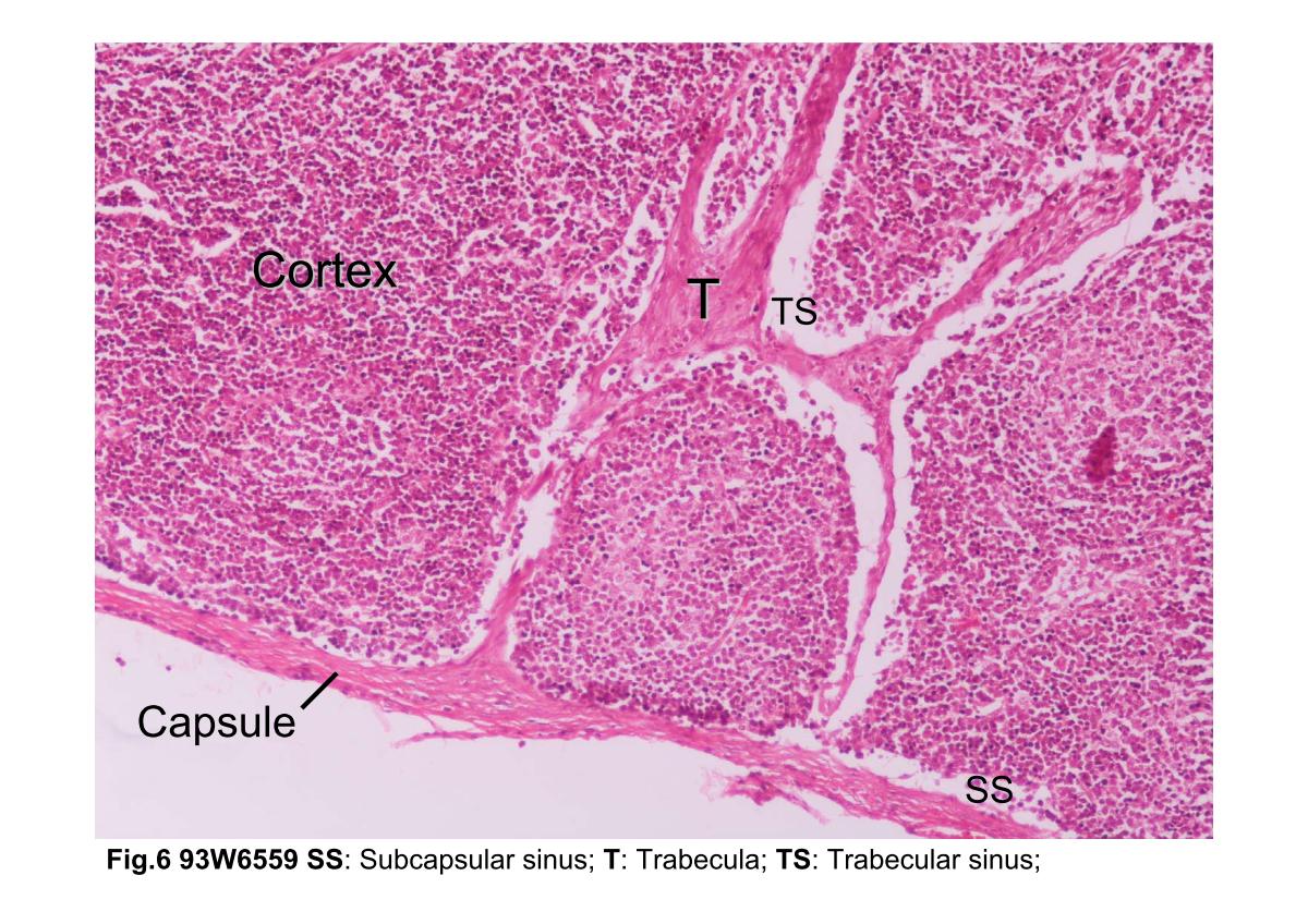
Fig.6 Photomicrograph of a lymphatic nodule. The capsule is composed of dense connective tissue from which trabeculae (T) penetrate into the organ. Immediately below the capsule is the subcapsular sinus (SS), which receives lymph from the afferent lymphatic vessels after they penetrate the capsule. The subcapsular sinus is continuous with the trabecular sinuses (TS) that course along the trabeculae (T).
block3_03block3_04block3_06block3_08block3_10block3_12block3_14block3_16block3_18block3_20block3_22

支援訊息