Up Block2 Prev Next

 上一張相片  下一張相片  索引頁
block3_22.jpg - Fig.11 93W4126 Thymus, H&E. Examination of the thymus atlow magnification reveals the lobules separated by adiposetissue (Ad). In the older thymus, much adipose tissue ispresent between the lobules. Each lobule is composed of adark-staining basophilic cortex (C) and a lighter-stainingmedulla (M). The cortex contains numerous densely packedlymphocytes, whereas the medulla contains fewerlymphocytes and is consequently less densely. The lobulesare not completely separate units; rather, they areinterconnected.

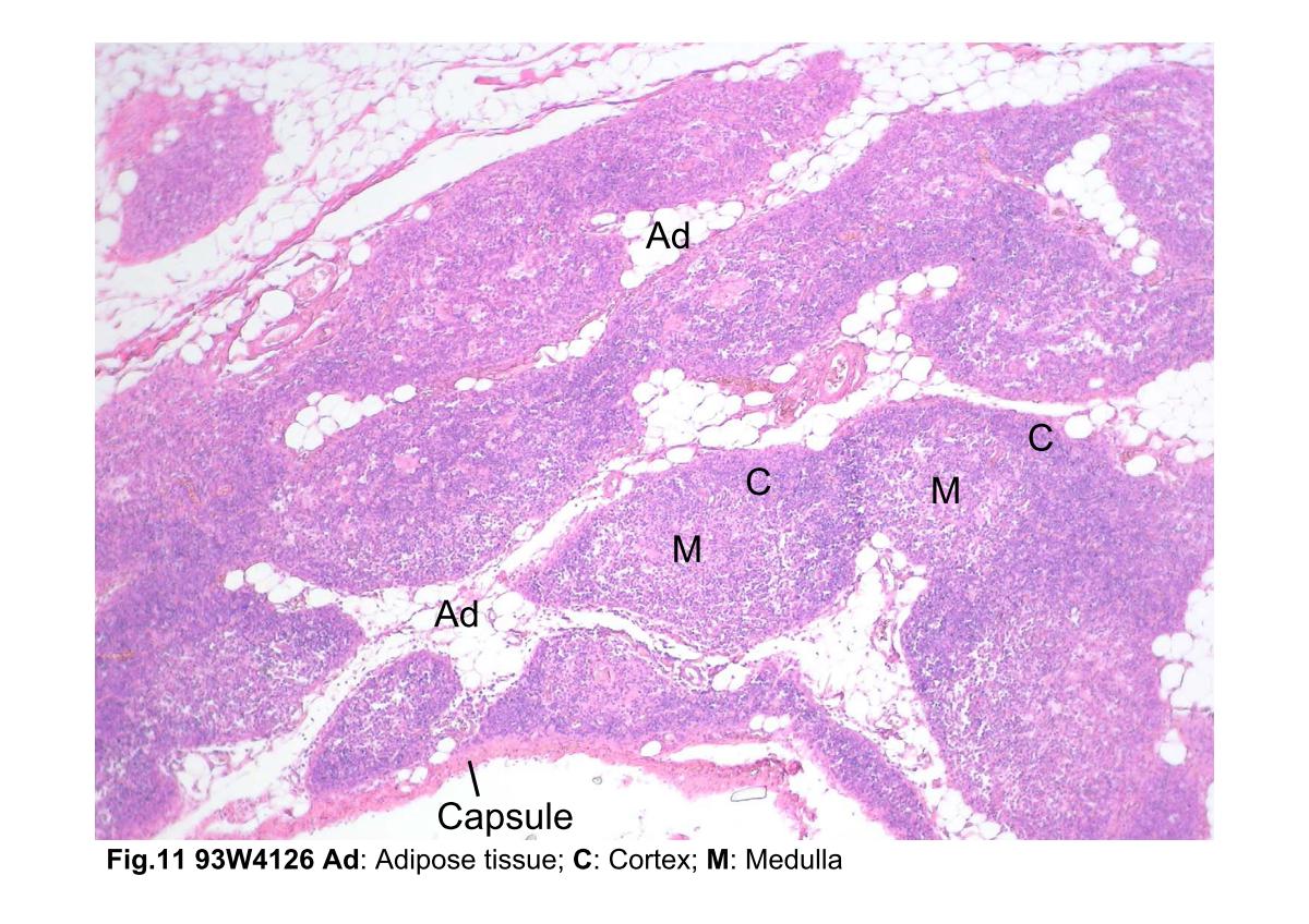
Fig.11 93W4126 Thymus, H&E. Examination of the thymus at low magnification reveals the lobules separated by adipose tissue (Ad). In the older thymus, much adipose tissue is present between the lobules. Each lobule is composed of a dark-staining basophilic cortex (C) and a lighter-staining medulla (M). The cortex contains numerous densely packed lymphocytes, whereas the medulla contains fewer lymphocytes and is consequently less densely. The lobules are not completely separate units; rather, they are interconnected.
block3_12block3_14block3_16block3_18block3_20block3_22block3_24block3_26block3_28block3_29 

支援訊息