Up Block2 Prev Next

 上一張相片  下一張相片  索引頁
block3_10.jpg - Fig.5 Photomicrograph of a lymphatic nodule. The capsuleis composed of dense connective tissue. Below the capsule isthe subcapsular sinus (SS). The area (black dashed line)shows the lymphatic nodule with a pale germinal center (GC)and a darker stained mantle zone (MZ) surrounding it. B cellsproliferate and differentiate in the germinal centers.

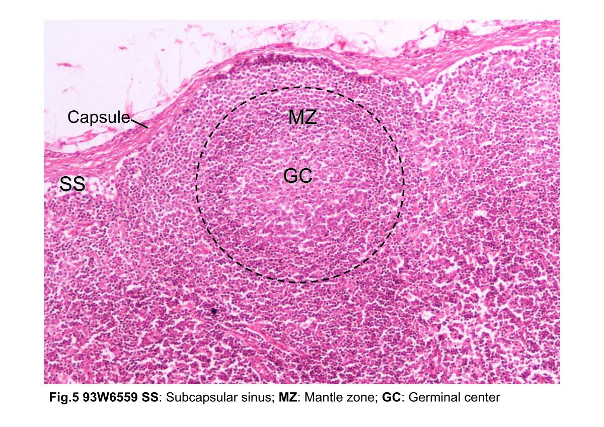
Fig.5 Photomicrograph of a lymphatic nodule. The capsule is composed of dense connective tissue. Below the capsule is the subcapsular sinus (SS). The area (black dashed line) shows the lymphatic nodule with a pale germinal center (GC) and a darker stained mantle zone (MZ) surrounding it. B cells proliferate and differentiate in the germinal centers.
block3_02block3_03block3_04block3_06block3_08block3_10block3_12block3_14block3_16block3_18block3_20

支援訊息