Up Block1 Prev Next

 上一張相片  下一張相片  索引頁
block1_14.jpg - Fig 4. The microscopic structure of trachea.The lining of the trachea consists of pseudostratified ciliatedcolumnar epithelium. Although the epithelium appears to formthe stratified, but all the cells rest on the basement membrane.The wall of the trachea contains the C-shaped tracheal cartilage.Besides, the tracheal cartilage is surrounded by the regulardense regular connective tissue (DCT). The collagen fibers arearranged in a regular manner and the fibroblast nuclei areelongated in the direction of the collagen fibers. By the way,adipose tissue (Ad) and nerve fiber bundles (NB) could be seenin this micrograph. Pseudostratified ciliated columnarepithelium appears at higher magnification in Fig 6b, and thecilia (C) is well demonstrated in this figure.

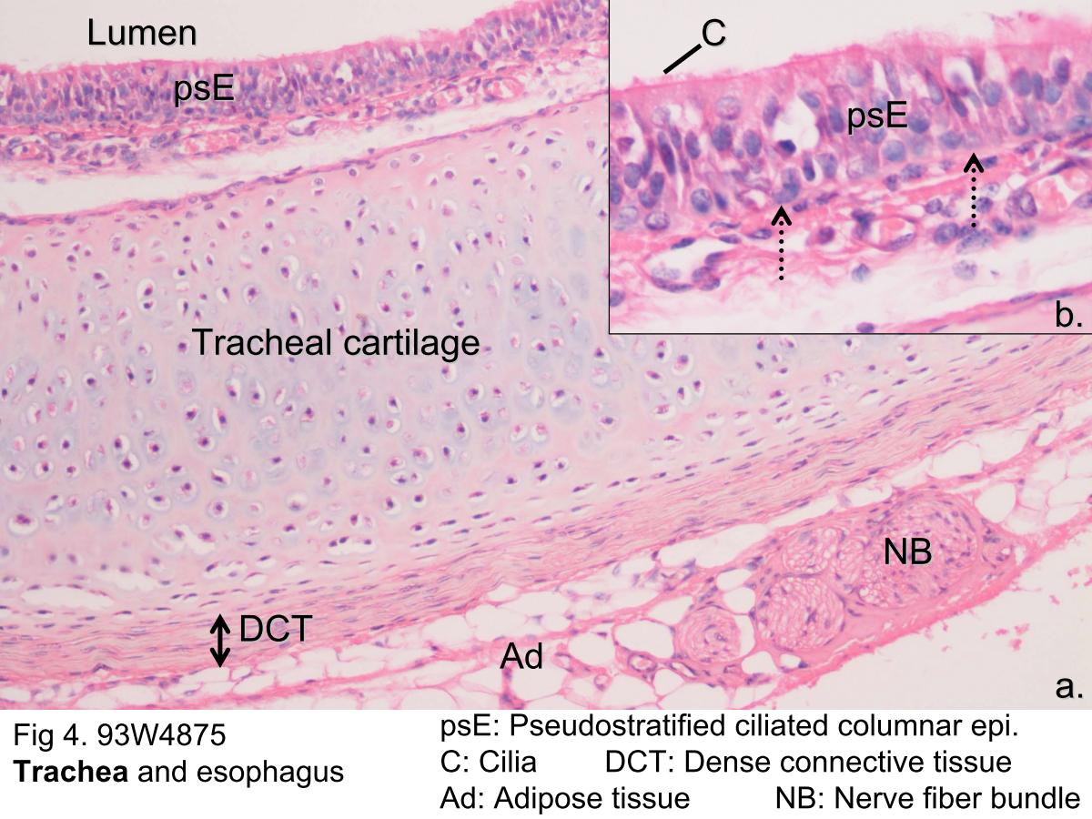
Fig 4. The microscopic structure of trachea. The lining of the trachea consists of pseudostratified ciliated columnar epithelium. Although the epithelium appears to form the stratified, but all the cells rest on the basement membrane. The wall of the trachea contains the C-shaped tracheal cartilage. Besides, the tracheal cartilage is surrounded by the regular dense regular connective tissue (DCT). The collagen fibers are arranged in a regular manner and the fibroblast nuclei are elongated in the direction of the collagen fibers. By the way, adipose tissue (Ad) and nerve fiber bundles (NB) could be seen in this micrograph. Pseudostratified ciliated columnar epithelium appears at higher magnification in Fig 6b, and the cilia (C) is well demonstrated in this figure.
block1_02block1_05block1_06block1_07block1_08block1_10block1_12block1_14block1_16block1_18block1_20block1_22block1_24block1_26block1_28

支援訊息