|
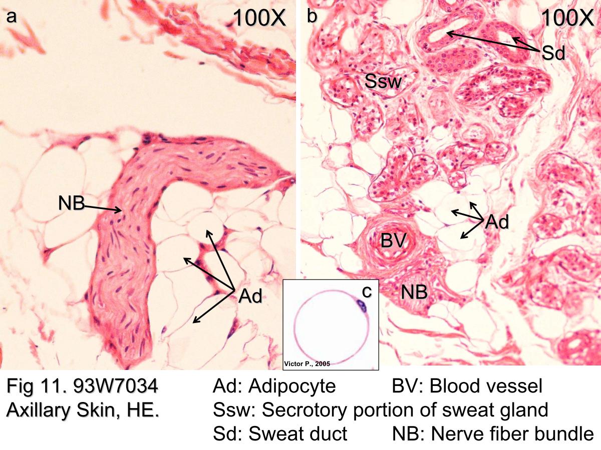 |
| Fig 11. Hypodermis of the thick skin. The lower magnification photomicrograph shows part of the hypodermis of the thick skin. It contains abundant adipocytes. The adipocyte (Ad) nucleus is compressed and displaced to one side of the stored lipid droplets and the cytoplasm including organelles is reduced to a small rim (Fig 11c). Fig 11a shows several adipocytes and nerve fiber bundles (NB). Fig 11b shows profiles of an eccrine sweat gland (Sw) and several blood vessels (BV). The cross sections of a blood vessel and a sweat gland can also be seen. However, the wall of blood vessels is composed of the squamous epithelium and the smooth muscle layer. The sweat gland has two parts: the secretory portion (Ssw) and the excretory duct (sweat duct, Sd). The secretory cells are arranged in circles (pseudostratified epithelium) having small lumen and the wall of the sweat duct is composed of stratified cuboidal cells. | |||||||||||||||
支援訊息