Up Block1 Prev Next

 上一張相片  下一張相片  索引頁
block1_16.jpg - Fig 5. 93W3039 Transitional epithelium, H&E. Thismicrograph is taken from the cross-sectioned ureter. The lumenof the ureter is lined by transitional epithelium (TE). The uretersare muscular tubes that carry urine from the kidneys to thebladder. The wall of the ureter contains smooth muscle (Sm).Because of contraction of the smooth muscle, the luminalsurface is characteristically folded, thus creating a star-likelumen. Surrounding the muscular wall is a loose connectivetissue containing adipose tissue (Ad) and blood vessels (BV).The wall of the ureter in the rectangular area in Fig 7a isexamined at higher magnification as in Fig 7b. Adipose tissue(Ad), smooth muscle (Sm) and blood vessels (BV) could beseen in this micrograph.

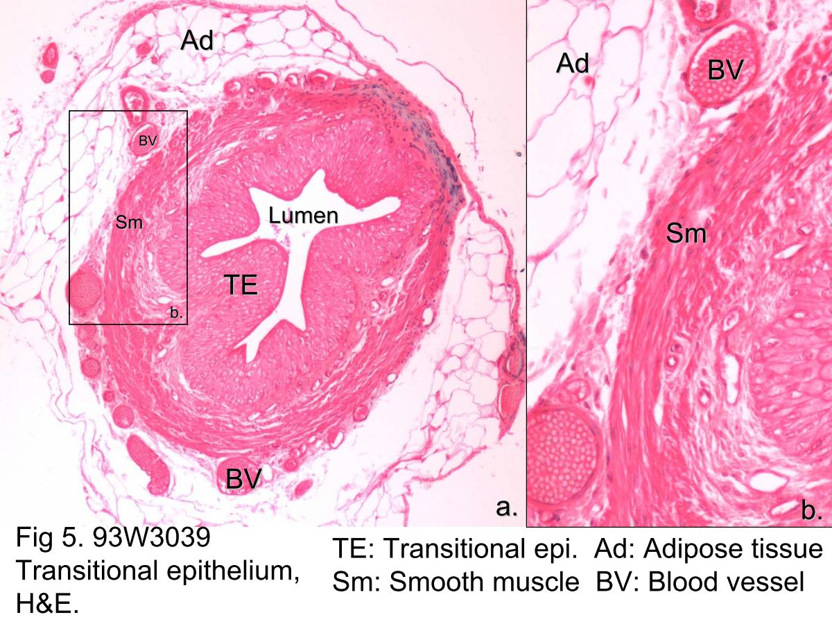
Fig 5. 93W3039 Transitional epithelium, H&E. This micrograph is taken from the cross-sectioned ureter. The lumen of the ureter is lined by transitional epithelium (TE). The ureters are muscular tubes that carry urine from the kidneys to the bladder. The wall of the ureter contains smooth muscle (Sm). Because of contraction of the smooth muscle, the luminal surface is characteristically folded, thus creating a star-like lumen. Surrounding the muscular wall is a loose connective tissue containing adipose tissue (Ad) and blood vessels (BV). The wall of the ureter in the rectangular area in Fig 7a is examined at higher magnification as in Fig 7b. Adipose tissue (Ad), smooth muscle (Sm) and blood vessels (BV) could be seen in this micrograph.
block1_05block1_06block1_07block1_08block1_10block1_12block1_14block1_16block1_18block1_20block1_22block1_24block1_26block1_28block1_30

支援訊息