Up Block1 Prev Next

 上一張相片  下一張相片  索引頁
block1_12.jpg - Fig 3. The microscopic structure of esophagus.The lining of the esophagus consists of more than one layer ofcells, and the surface layer consists of flat or squamous cell. It'sa good example of stratified squamous epithelium (SS). In astratified epithelium, the shape and height of the cells usuallyvary from layer to layer, but only the shape of the cells that formthe surface layer is used in classifying the epithelium. The wallof the esophagus contains the smooth muscle and the skeletalmuscle. You could try to discriminate smooth muscle (cross-sectioned in this micrograph) from skeletal muscle (longitudinal-sectioned in this micrograph) at low-magnification immediately.By the way, the cross-striation of the skeletal muscle appear athigher magnification in Fig 5b.

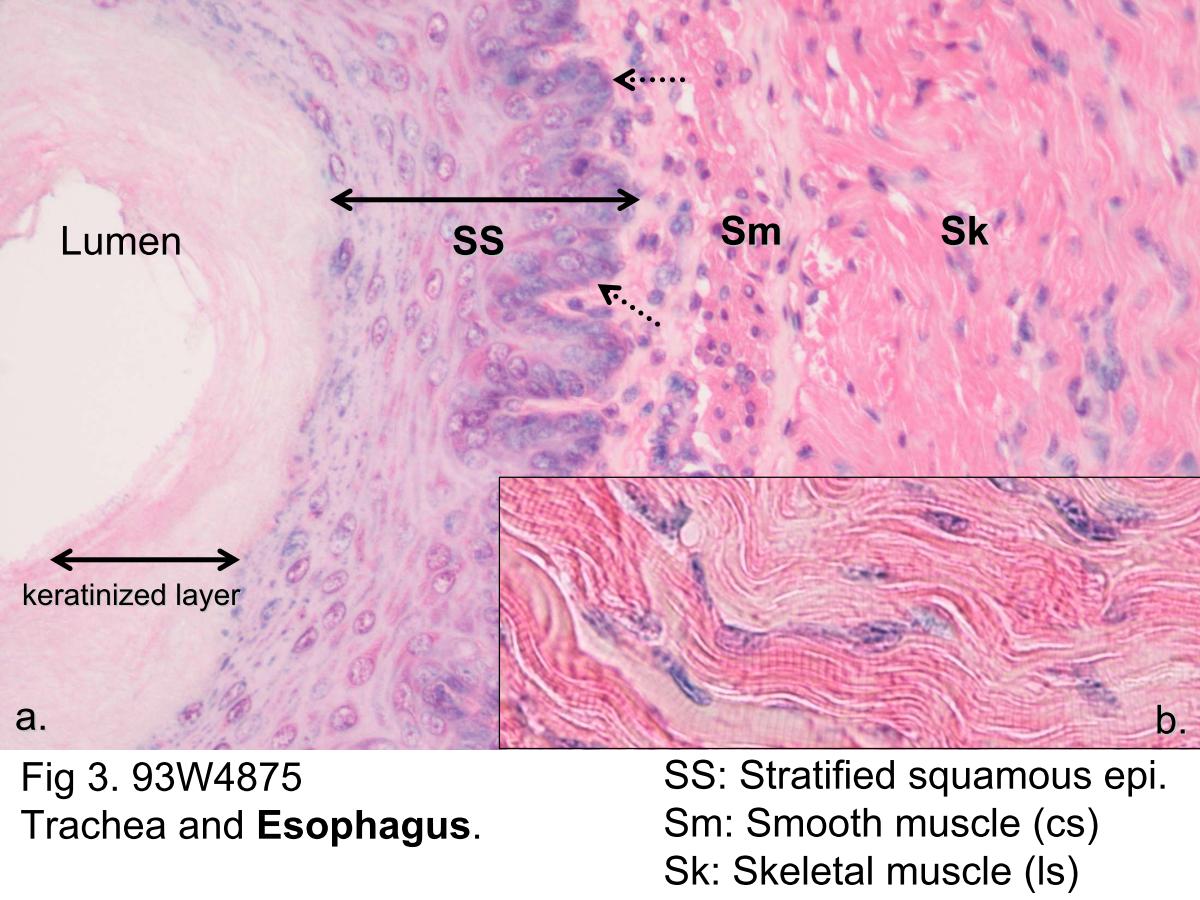
Fig 3. The microscopic structure of esophagus. The lining of the esophagus consists of more than one layer of cells, and the surface layer consists of flat or squamous cell. It's a good example of stratified squamous epithelium (SS). In a stratified epithelium, the shape and height of the cells usually vary from layer to layer, but only the shape of the cells that form the surface layer is used in classifying the epithelium. The wall of the esophagus contains the smooth muscle and the skeletal muscle. You could try to discriminate smooth muscle (cross- sectioned in this micrograph) from skeletal muscle (longitudinal- sectioned in this micrograph) at low-magnification immediately. By the way, the cross-striation of the skeletal muscle appear at higher magnification in Fig 5b.
block1_01block1_02block1_05block1_06block1_07block1_08block1_10block1_12block1_14block1_16block1_18block1_20block1_22block1_24block1_26

支援訊息