|
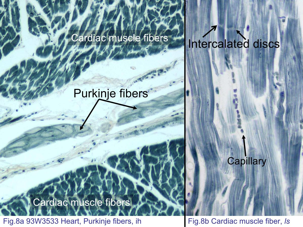 |
| Fig.8a 93W3533 Heart, Purkinje fibers, ih. Purkinje fibers
are the components of the heart¡¦s conduction system. They
are more easily observed in a slide stained with iron
hematoxylin. While most common in the subendothelial space,
they can also be observed in the myocardium. Compare
Purkinje fibers with cardiac muscle fibers. Note larger
numbers of myofibrils and smaller cell size in cardiac muscle
fibers. The Purkinje fibers contain large amounts of invisible
glycogen particles, which appear as pale-staining regions that
occupy the center portion of the cell surrounded by the
myofibrils. Fig.8b Cardiac muscle fiber, ls. Iron hematoxylin staining of cardiac muscle highlights the cross striations and intercalated discs. The red blood cells are stained in black dots. The capillary diameter is about the same size as a red blood cell. In longitudinal section, the capillaries will look to appear as rows of red blood cells. |
|||||||||||
支援訊息