Up Block3 Prev Next

 上一張相片  下一張相片  索引頁
block4_08.jpg - Fig.4 Photomicrograph of pulmonary valve. The heartvalves consist of fibroelastic tissue, the lamina fibrosa. Thesurfaces covered by a thin layer of endothelium continuouswith the lining of both the heart chambers and great vessels.This micrograph shows the pulmonary valve arising at thejunction of the walls of the right ventricle and the pulmonarytrunk.

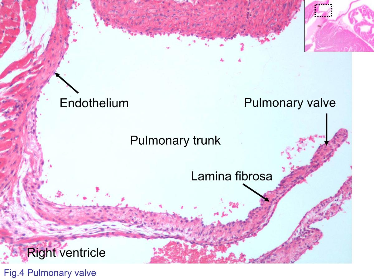
Fig.4 Photomicrograph of pulmonary valve. The heart valves consist of fibroelastic tissue, the lamina fibrosa. The surfaces covered by a thin layer of endothelium continuous with the lining of both the heart chambers and great vessels. This micrograph shows the pulmonary valve arising at the junction of the walls of the right ventricle and the pulmonary trunk.
block4_01block4_02block4_03block4_04block4_06block4_08block4_10block4_12block4_14block4_16block4_18

支援訊息