|
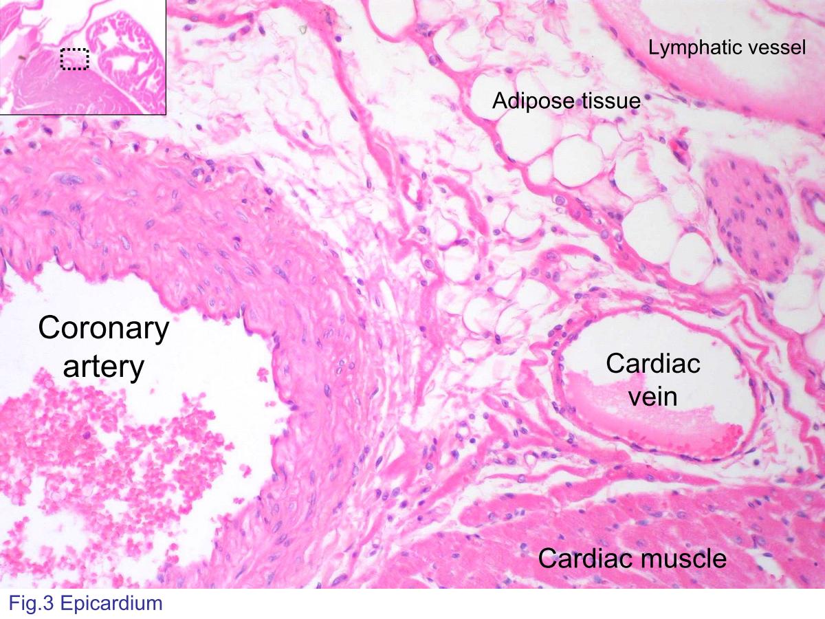 |
| Fig.3 Photomicrograph of epicardium (visceral
pericardium). Here shows an area where the epicardium
contains the branches of coronary artery and the cardiac vein.
There is a lymphatic vessel without blood cells inside in this
slide. The black dashed line rectangle in inset figure shows the orientation of the photomicrograph in lower magnification. No more explanation is given in the following figures. |
|||||||||||
支援訊息