Up Block3 Prev Next

 上一張相片  下一張相片  索引頁
block4_20.jpg - Fig.10 93W6515 Artery cs v&e. This slide is stained withVerhoeff's stain to visualize the elastic fibers, and with eosin toshow the cellular structures. Muscular arteries have moresmooth muscle and less elastin in the tunica media thanelastic arteries. The muscular arteries are characterized by alayer of internal elastic lamina separating the tunica intimafrom the tunica media. The less prominent and more variableexternal elastic lamina lies between the tunica media and theadventitia. The tunica intima consists of an endothelial liningand a small amount of connective tissue. However, nucleus ofthe endothelial cells can¡¦t be observed by the v&e stainingmethod. The tunica adventitia is composed of collagen fibers(pink), elastic fibers (black) and vasa vasorum.

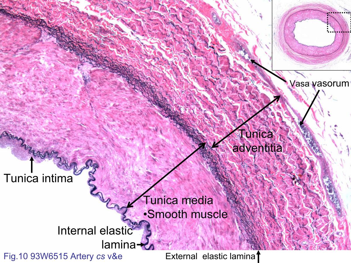
Fig.10 93W6515 Artery cs v&e. This slide is stained with Verhoeff's stain to visualize the elastic fibers, and with eosin to show the cellular structures. Muscular arteries have more smooth muscle and less elastin in the tunica media than elastic arteries. The muscular arteries are characterized by a layer of internal elastic lamina separating the tunica intima from the tunica media. The less prominent and more variable external elastic lamina lies between the tunica media and the adventitia. The tunica intima consists of an endothelial lining and a small amount of connective tissue. However, nucleus of the endothelial cells can¡¦t be observed by the v&e staining method. The tunica adventitia is composed of collagen fibers (pink), elastic fibers (black) and vasa vasorum.
block4_10block4_12block4_14block4_16block4_18block4_20block4_22block4_24block4_26block4_27block4_28

支援訊息