|
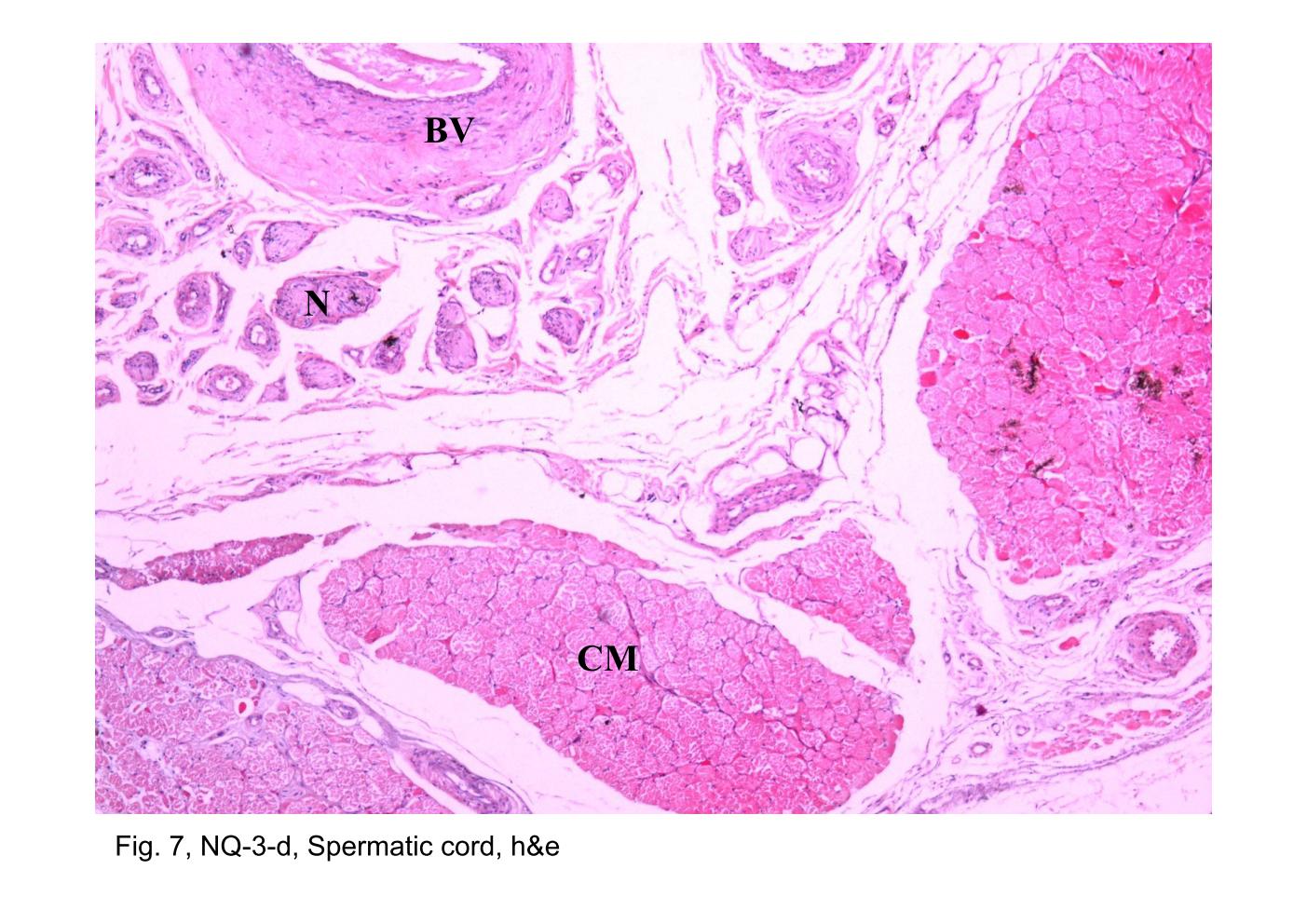 |
| Fig. 6 & 7, NQ-3-d, Spermatic cord, h&e
The ductus deferens and some of the blood vessels (BV), nerves (N) and
cremaster muscles (CM) accompaning the duct in the spermatic cord are
shown in this cross section. The wall of the ductus deferens is extremely
thick, mostly because of the presence of abundent smooth muscle. During
the preparation of the tissue, The muscle contracted and caused the
mucosa to form longitudinal folds. For this reason, the lumen (L) usually
appears irregular in the section. The smooth muscle of the ductus deferens is arranged as a thick outer longitudinal layer (OL), a thick middle circular layer (MC), and a thin inner longitudinal layer (IL). The epithelial lining of the ductus deferens consists of the pseudostratified columnar epithelium (PCE). Between the epithelium and the inner longitudinal smooth muscle layer, there is a cellular layer of loose connective tissue, the lamina propria (LP). |
|||||||||||
支援訊息