|
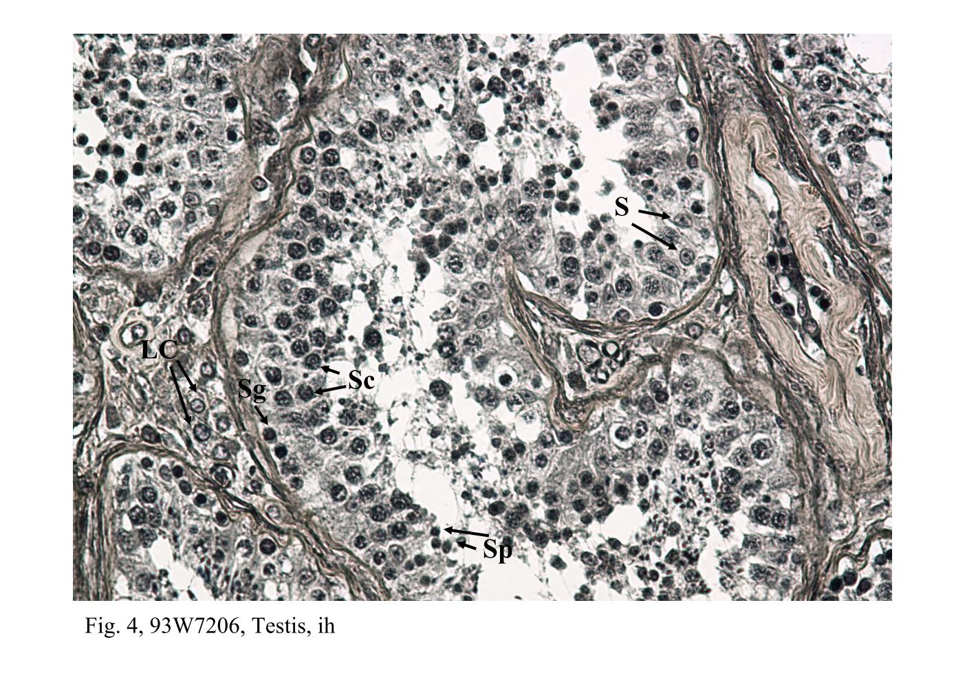 |
| Fig. 3 & 4, Q-1-b, Testis, h&e; 93W7206, Testis, ih
Examination of the tubule epithelium reveals two kinds of cells: a
proliferating population of spermatogenic cells and a non-proliferating
population, the Sertoli cells (S). The Sertoli cells are considerably fewer
and can be recognized by their elongate, trangular, pale-staining nuclei
and conspicuous nucleolus. The Sertoli cell extends from the periphery of
the tubule to the lumen. The spermatogenic cells consist of successive
generations arranged in concentric layers. The spermatogonia (Sg) are
found at the periphery. The spermatocytes (Sc), most of them have large
round nuclei with a distinctive chromatin pattern, come to lie above the
spermatogonia. The spermatid (Sp) consists of one or two generations and
occupies the site closest to the lumen. In high magnification, it reveals a population of Leydig cells (LC) that occur in small clusters and lie in the interstitial space between adjacent tubules. They are readily identified by their location, small round nucleus and eosinophilic cytoplasm. |
|||||||||||
支援訊息