|
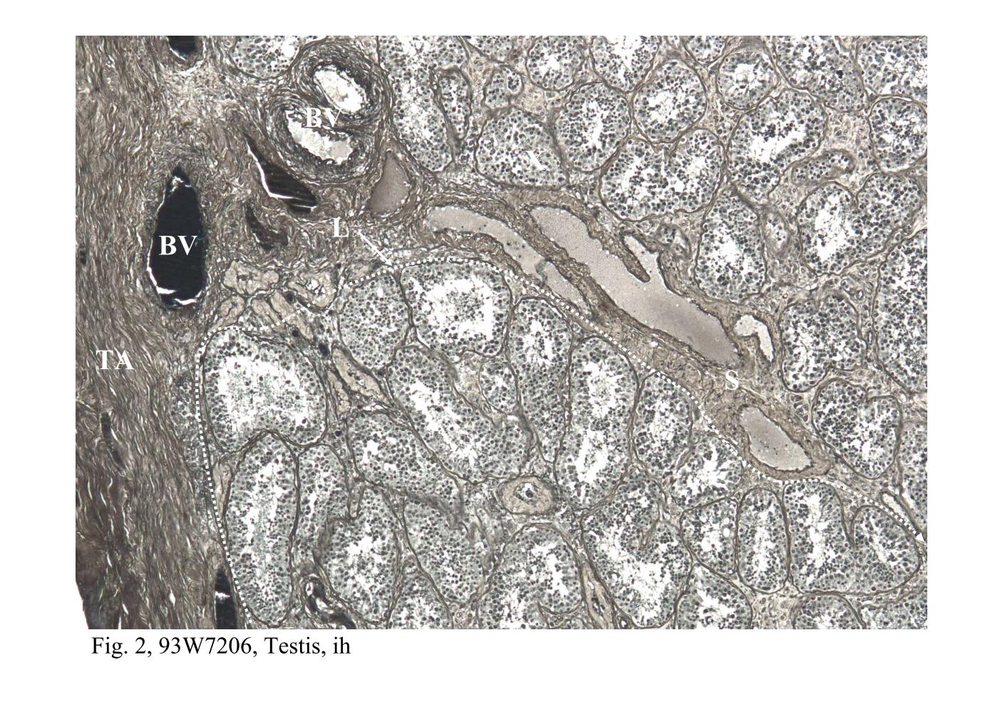 |
| Fig. 1 & 2, Q-1-b, Testis, h&e; 93W7206, Testis, ih
The seminiferous tubules and the tunica albuginea (TA or capsule) of the testis
organ are identified in this section. Extending from the capsule are connective
tissue septa (S) that divide the organ into several ( about 250) compartments or
lobules. Each compartment (L: corresponding to a lobule) contains coiled
seminiferous tubules. Blood vessels (BV) are abundant under the capsule that
located between the tunica albuginea and seminiferous tubules is referred as the
tunica vasculosa. The branches of the blood vessels extending within the
connective tissue septa are also observed. The seminiferous tubules are convoluted, therefore, the profiles of them present in a section are variable in appearance. Sometimes, the wall of a tubule is sectioned tangentially, thus obscuring the lumen and revealing what appears to be a solid mass of cells (X). |
|||||||||||
支援訊息