Up Block6-2 Prev Next

 上一張相片  下一張相片  索引頁
block6-2_13.jpg - Fig 5. 93W6748 esophagus, middle region (cs) H&E.Fig 5a. The muscularis externa (M) of the middle portion of theesophagus is composed of smooth muscle and skeletal muscle.The muscle fibers are arranged into two layers (inner circular &outer longitudinal).Smooth muscle cells is spindle-shaped in the longitudinalsection (Fig 5b). Their nuclei (N) are also elongated andconform to the general shape of the cell. A nuclei display slightlytwisted is indicated by an arrow, like a corkscrew; this ischaracteristic of contracted cells.Fig 5c shows a cross-sectioned of the smooth muscle cells,displaying circular or polygonal profiles with variations on size.In most of the cells, the nuclei (N) have not been included in thesection, and only the eosinophilic cytoplasm appears.

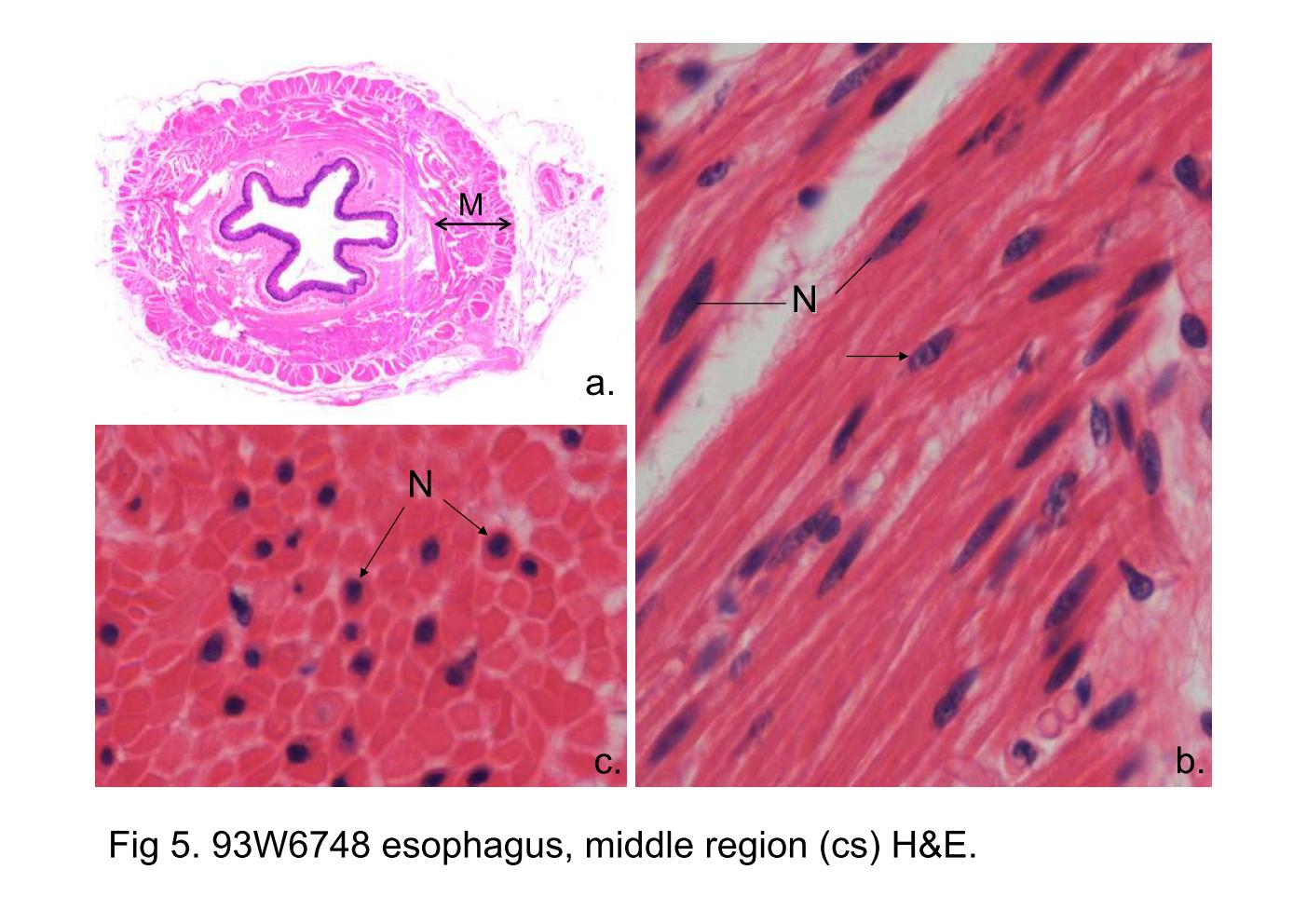
Fig 5. 93W6748 esophagus, middle region (cs) H&E.

Fig 5a. The muscularis externa (M) of the middle portion of the esophagus is composed of smooth muscle and skeletal muscle. The muscle fibers are arranged into two layers (inner circular & outer longitudinal).

Smooth muscle cells is spindle-shaped in the longitudinal section (Fig 5b). Their nuclei (N) are also elongated and conform to the general shape of the cell. A nuclei display slightly twisted is indicated by an arrow, like a corkscrew; this is characteristic of contracted cells.

Fig 5c shows a cross-sectioned of the smooth muscle cells, displaying circular or polygonal profiles with variations on size. In most of the cells, the nuclei (N) have not been included in the section, and only the eosinophilic cytoplasm appears.

block6-2_04block6-2_05block6-2_07block6-2_09block6-2_11block6-2_13block6-2_15    

支援訊息