Up Block6-2 Prev Next

 上一張相片  下一張相片  索引頁
block6-2_07.jpg - Fig 2. 93W6746 Esophagus, upper region (c.s.) H&E.Fig 2a. The muscularis externa (M) of the upper portion of theesophagus belongs to the skeletal muscle, appearing the innercircular and outer longitudinal arrangement patterns.Fig 2b shows high-magnification of cross sectioned musclecells, appearing the polygonal profiles. Notice the numerous ofperipherally located multinuclei (N). Endomysium (E) is a layerof basal lamina, surrounding each individual muscle fibers.            Skeletal muscle

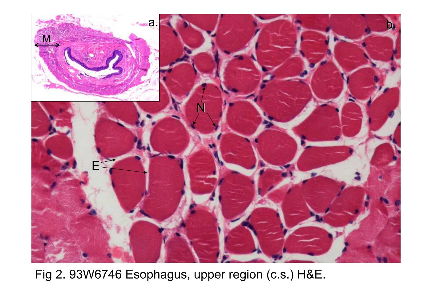
Fig 2. 93W6746 Esophagus, upper region (c.s.) H&E.

Fig 2a. The muscularis externa (M) of the upper portion of the esophagus belongs to the skeletal muscle, appearing the inner circular and outer longitudinal arrangement patterns.

Fig 2b shows high-magnification of cross sectioned muscle cells, appearing the polygonal profiles. Notice the numerous of peripherally located multinuclei (N). Endomysium (E) is a layer of basal lamina, surrounding each individual muscle fibers. Skeletal muscle

block6-2_01block6-2_02block6-2_03block6-2_04block6-2_05block6-2_07block6-2_09block6-2_11block6-2_13block6-2_15 

支援訊息