Up Block6-2 Prev Next

 上一張相片  下一張相片  索引頁
block6-2_11.jpg - Fig 4. 93W6245 Cardiac Muscle (cs&ls) ih.Fig 4a shows a longitudinal section of cardiac muscle fibers.Cardiac muscle fiber is cylindrical profile and arranged end toend, connecting by specialized cell junctions, intercalated discs(ID). Intercalated disk provide the site where it can join two ormore cardiac fibres, creating the branched patterns (arrow). Theovoid nuclei (N) are centrally located and the perinuclear spaceis devoid of myofibrils, showing pale staining in the cross-sectionof cardiac muscle (Fig 4b, asterisks).

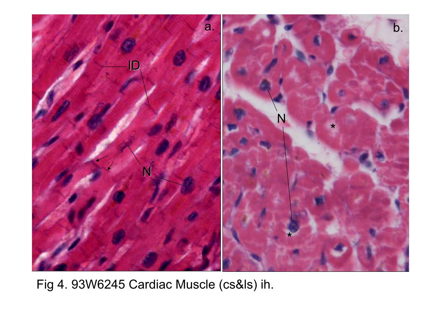
Fig 4. 93W6245 Cardiac Muscle (cs&ls) ih. Fig 4a shows a longitudinal section of cardiac muscle fibers. Cardiac muscle fiber is cylindrical profile and arranged end to end, connecting by specialized cell junctions, intercalated discs (ID). Intercalated disk provide the site where it can join two or more cardiac fibres, creating the branched patterns (arrow). The ovoid nuclei (N) are centrally located and the perinuclear space is devoid of myofibrils, showing pale staining in the cross-section of cardiac muscle (Fig 4b, asterisks).
block6-2_03block6-2_04block6-2_05block6-2_07block6-2_09block6-2_11block6-2_13block6-2_15   

支援訊息