|
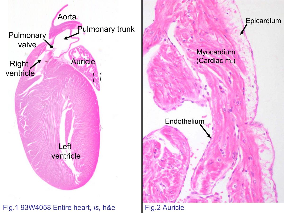 |
| Fig.1 93W4058 Entire heart, ls, h&e. Put the slide first on a
white paper to observe the overview orientation of the section.
However, every section of the slides is slightly different.
Compare your slide with this figure and identify the structures
in your slide. Fig.2 Photomicrograph of auricle. The auricle is easier to demonstrate the three-layered structure of the heart wall. The endocardium has a inner surface layer of flattened endo- thelium. The subendothelial tissue merges with the collagen fibers surrounding outer cardiac muscle (myocardium). The outermost epicardium is composed of connective tissue and the superficial mesothelial cells (not clearly seen here) which produce the pericardial fluid. |
|||||||||||
支援訊息