Up Block14 Prev Next

 上一張相片  下一張相片  索引頁
block14_49.jpg - Fig. 25, 93W7334, Vagina (ls), H&EThe mucosa of the vagina consists of a stratified squamous epithelium (SSE)   and an underlying fibrous connective tissue (CT) that often appears more   cellular than other fibrous connective tissue. The boundary between the   two is readily identified because of the conspicuous staining of the   closely packed small cells of the basal layer (B) of the epithelium.   Connective tissue papillae (P) project into the underside of the epithelium,   giving the epithelial-connective tissue junction an uneven appearance.   The papillae may be cut obliquely or in cross section and thus may   appear as connective tissue islands (arrows; insert). The epithelium is   characteristically thick and although keratohyaline granules may be   found in the superficial cells, keratinization does not occur in human   vaginal epithelium. Thus, nuclei can be observed throughout the entire   thickness of the epithelium despite the fact that the cytoplasm of most of   the cells above the basal layers appears empty. These cells are normally   filled with large deposits of glycogen that is lost in the processes of   fixation and embedding of the tissue.

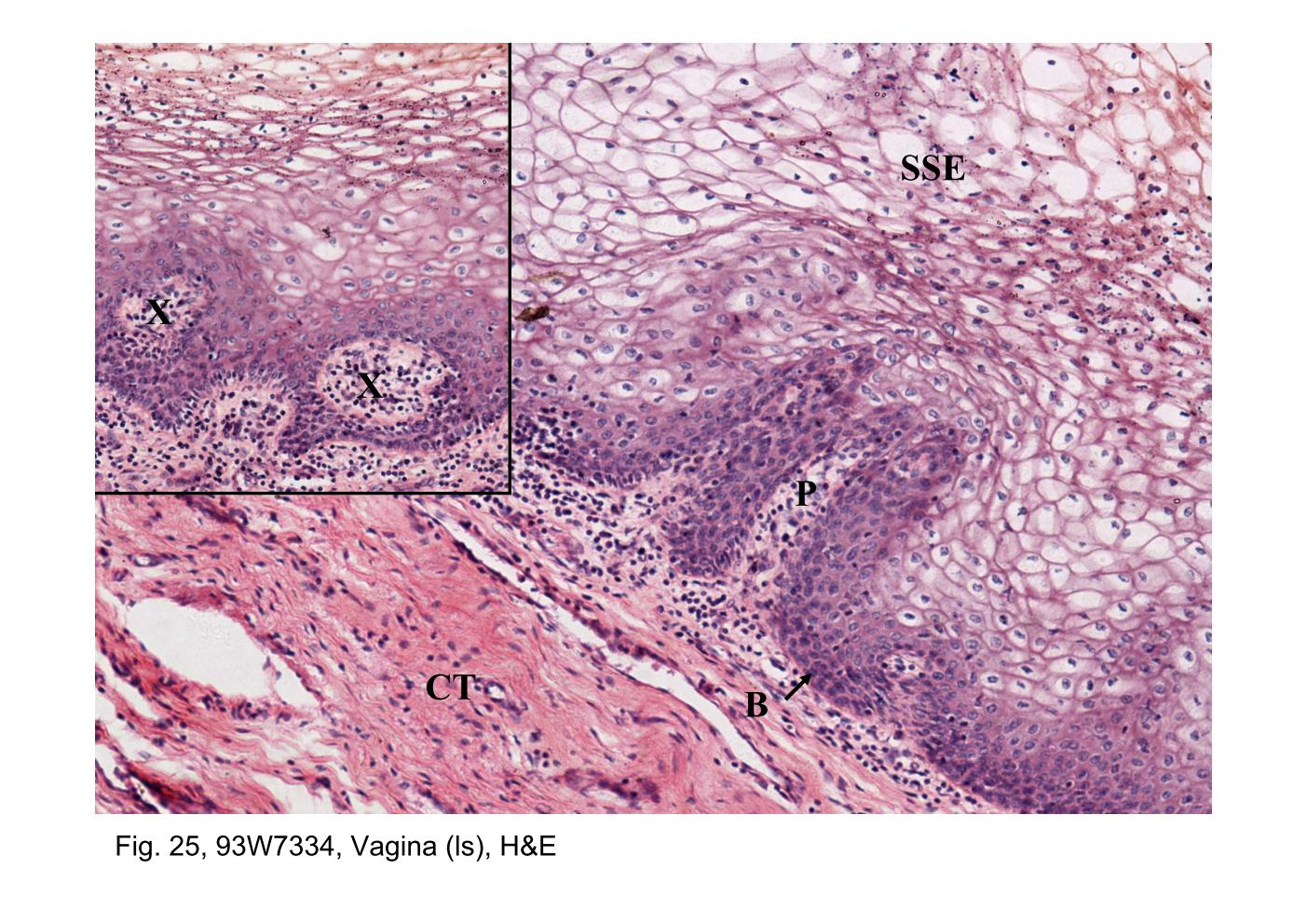
Fig. 25, 93W7334, Vagina (ls), H&E The mucosa of the vagina consists of a stratified squamous epithelium (SSE) and an underlying fibrous connective tissue (CT) that often appears more cellular than other fibrous connective tissue. The boundary between the two is readily identified because of the conspicuous staining of the closely packed small cells of the basal layer (B) of the epithelium. Connective tissue papillae (P) project into the underside of the epithelium, giving the epithelial-connective tissue junction an uneven appearance. The papillae may be cut obliquely or in cross section and thus may appear as connective tissue islands (arrows; insert). The epithelium is characteristically thick and although keratohyaline granules may be found in the superficial cells, keratinization does not occur in human vaginal epithelium. Thus, nuclei can be observed throughout the entire thickness of the epithelium despite the fact that the cytoplasm of most of the cells above the basal layers appears empty. These cells are normally filled with large deposits of glycogen that is lost in the processes of fixation and embedding of the tissue.
block14_39block14_41block14_43block14_45block14_47block14_49block14_51block14_53block14_55block14_57block14_59

支援訊息