Up Block10-2 Prev Next

 上一張相片  下一張相片  索引頁
block10-2_17.jpg - Fig 7. 93W4570, Liver, human (sec.) H&E.This figure focuses on the structure of the portal area. Portalarea is a connective tissue septa that carries the branches ofthe hepatic artery, the portal vein, and the bile duct. Thelargest tube is a terminal branch of the hepatic portal vein(HPV) which has very thin wall lined by flattened endothelialcells. Smaller diameter thick-walled vessels with the typicalstructure of arterioles are terminal branches of the hepaticartery (HA). The bile ducts (BD) are lined by simple cuboidalepithelium. They are usually located at the periphery of theportal area and are approximately the same size as thearterioles.

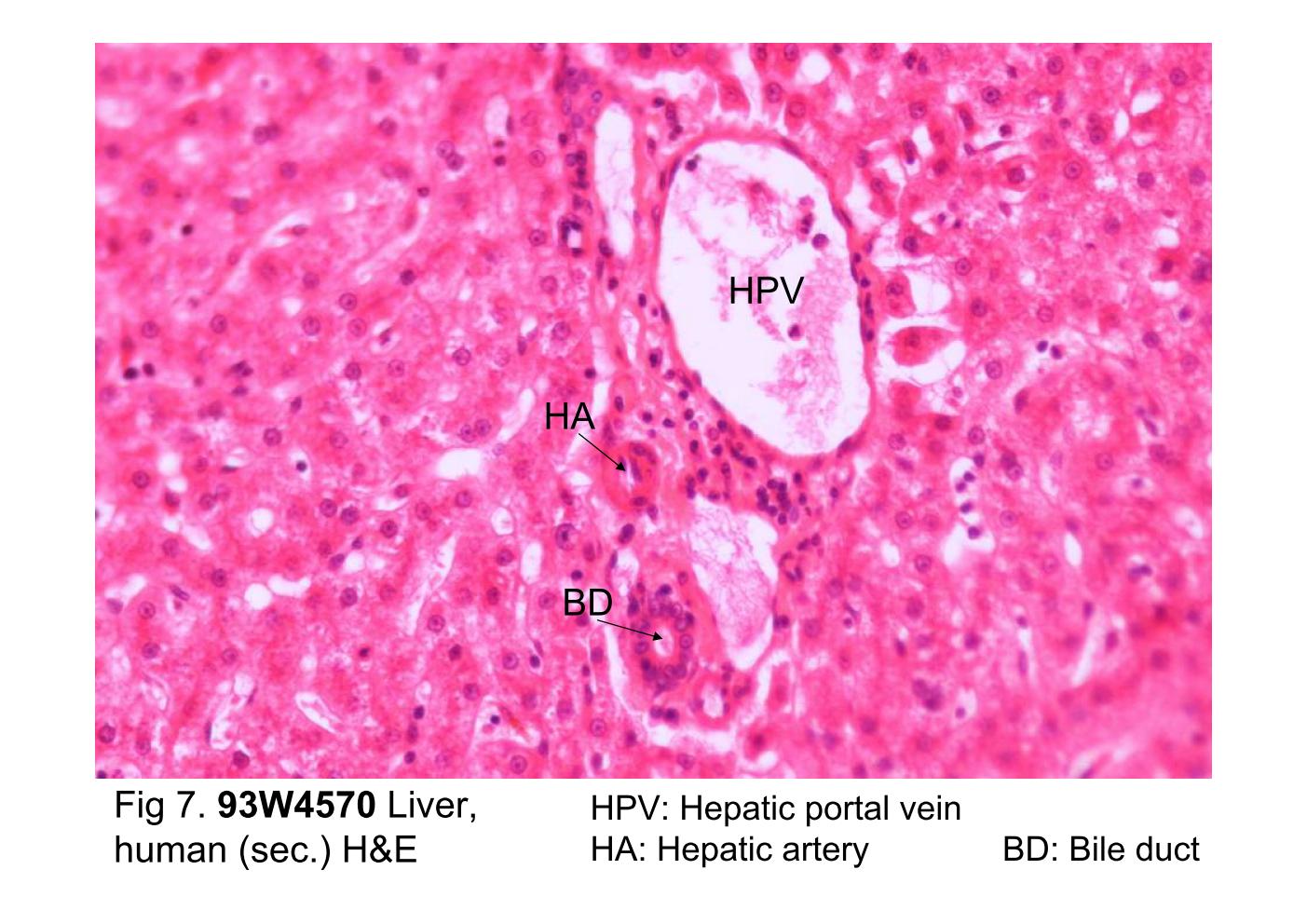
Fig 7. 93W4570, Liver, human (sec.) H&E. This figure focuses on the structure of the portal area. Portal area is a connective tissue septa that carries the branches of the hepatic artery, the portal vein, and the bile duct. The largest tube is a terminal branch of the hepatic portal vein (HPV) which has very thin wall lined by flattened endothelial cells. Smaller diameter thick-walled vessels with the typical structure of arterioles are terminal branches of the hepatic artery (HA). The bile ducts (BD) are lined by simple cuboidal epithelium. They are usually located at the periphery of the portal area and are approximately the same size as the arterioles.
block10-2_07block10-2_09block10-2_11block10-2_13block10-2_15block10-2_17block10-2_19block10-2_21block10-2_23block10-2_25block10-2_27

支援訊息