|
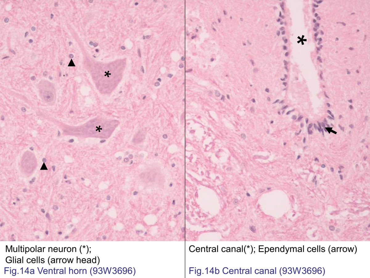 |
| Fig. 14a High magnification of ventral horn of spinal cord.
The ventral horn of the spinal cord contains large cell
bodies of multipolar motor neurons. The nuclei of neuroglial
cells are also evident, but their cytoplasm is not easily to
identified. Fig. 14b High magnification of ependymal cells. The ependymal cells are cuboid or columnar cell, arranging in a single layer pattern that is lining the central canal. |
|||||||||||
支援訊息