Up Block7 Prev Next

 上一張相片  下一張相片  索引頁
block7_26.jpg - Fig. 11 Photomicrograph of Meissner's corpuscles.Meissner's corpuscle housed in the apical of dermalpapillae and only lamellae cells of Meissner's corpusclecould be observed with the H&E staining (Fig. 11a).Meissner's corpuscle was innervated by nerve fibers thatdemonstrated with the pan-axonal (Fig. 11b).

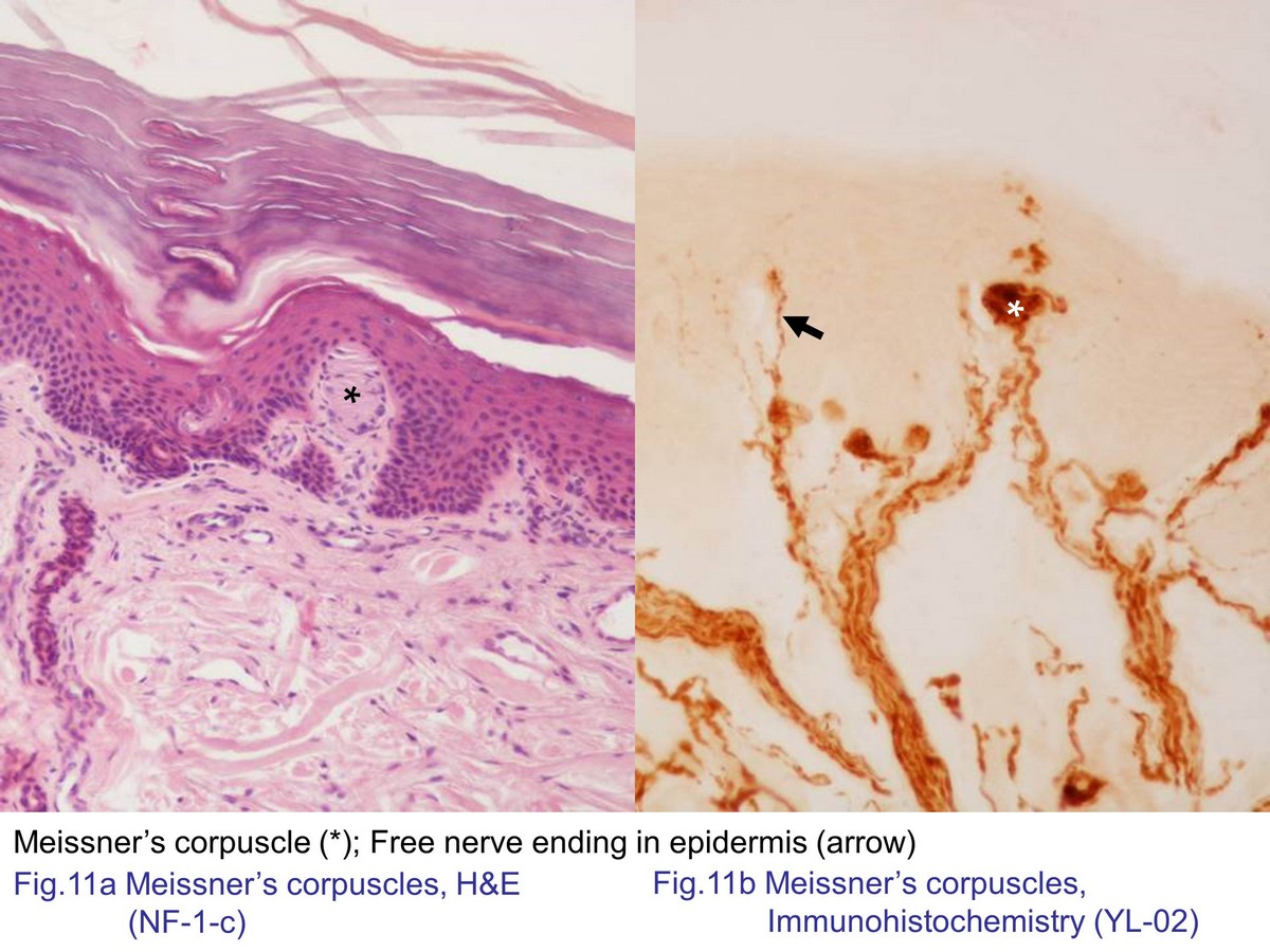
Fig. 11 Photomicrograph of Meissner's corpuscles. Meissner's corpuscle housed in the apical of dermal papillae and only lamellae cells of Meissner's corpuscle could be observed with the H&E staining (Fig. 11a). Meissner's corpuscle was innervated by nerve fibers that demonstrated with the pan-axonal (Fig. 11b).
block7_16block7_18block7_20block7_22block7_24block7_26block7_28block7_30block7_32block7_34block7_36

支援訊息