|
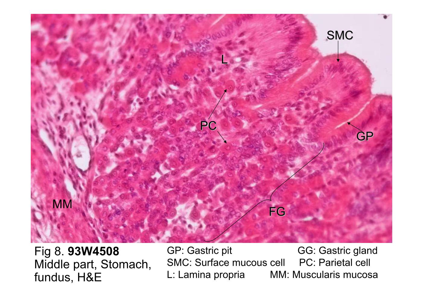 |
| Fig 8. 93W4508 Middle part, Stomach, fundus, H&E.
The gastric pit, the depression formed by the surface mucous
cells (SMC). Just below the gastric pits (GP) are the fundic
glands (FG), in which one can identify parietal cells (P). The
parietal cells are recognized by their copious eosinophilic
cytoplasm and central nucleus, which is often described as a
¡§fried egg appearance¡¨. The glands extend down to the
muscularis mucosa (MM). The lamina propria (L) is highly
cellular because of the presence of large numbers of
lymphocytes. Examine the cardiac and pylorus gland of this slide at higher magnification (not shown here). The cardiac glands are composed mainly of mucus-secreting cells. The pyloric glands are branched and coiled; the gastric pits of the pylorus occupy about half the thickness of the pyloric mucosa. |
|||||||||||
支援訊息