Up Block10-1 Prev Next

 上一張相片  下一張相片  索引頁
block10-1_25.jpg - Fig 11. 93W4523 Middle part, small intestine (sec.) H&E.Several cell types can be identified in this photomicrograph.Enterocytes (E) are tall columnar cells with surface microvillithat are seen as a striated border in light microscopy. Gobletcells (G) are scattered among the enterocytes and stainedpaler. The lamina propria (L) contains considerable numbers oflymphocytes and other cells of the immune system. Thelamina propria extends between the crypts and into the core ofeach villus and contains a rich vascular and lymphatic network.

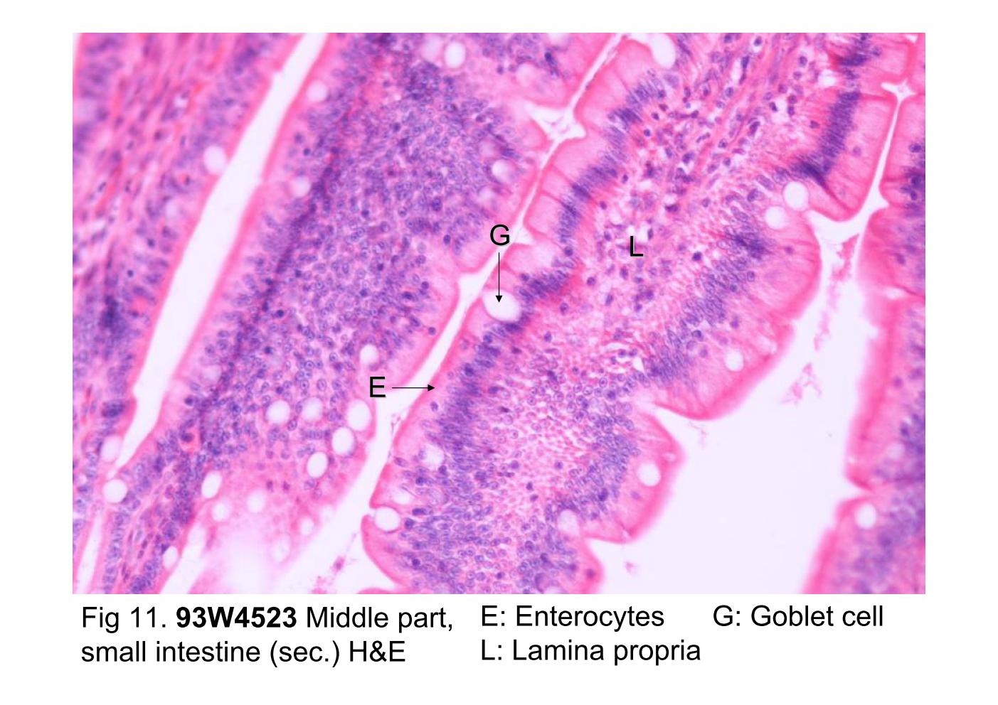
Fig 11. 93W4523 Middle part, small intestine (sec.) H&E. Several cell types can be identified in this photomicrograph. Enterocytes (E) are tall columnar cells with surface microvilli that are seen as a striated border in light microscopy. Goblet cells (G) are scattered among the enterocytes and stained paler. The lamina propria (L) contains considerable numbers of lymphocytes and other cells of the immune system. The lamina propria extends between the crypts and into the core of each villus and contains a rich vascular and lymphatic network.
block10-1_15block10-1_17block10-1_19block10-1_21block10-1_23block10-1_25block10-1_27block10-1_28   

支援訊息