Up Block10-1 Prev Next

 上一張相片  下一張相片  索引頁
block10-1_09.jpg - Fig 3. The microscopic structure of the muscularis externaof the esophagus.The muscularis externa consists of two muscle layers, an innercircular layer and an outer longitudinal layer. The esophagus isdivided histologically into three regions on the basis of the typeof muscle which muscularis externa contains. At the middle thirdof the esophagus, skeletal muscle (Sk) and smooth muscle (Sm)bundles are mixed and interwoven in the muscularis externa.Located between the two muscle layers is a thin connectivetissue layer. Within this connective tissue lies the Auerbach'splexus (Au) containing nerve cell bodies of postganglionicparasympathetic neurons and neurons of the enteric nervoussystem, as well as blood vessels and lymphatic vessels. Therectangle of Fig 3a is examined at higher magnification in Fig 3b.

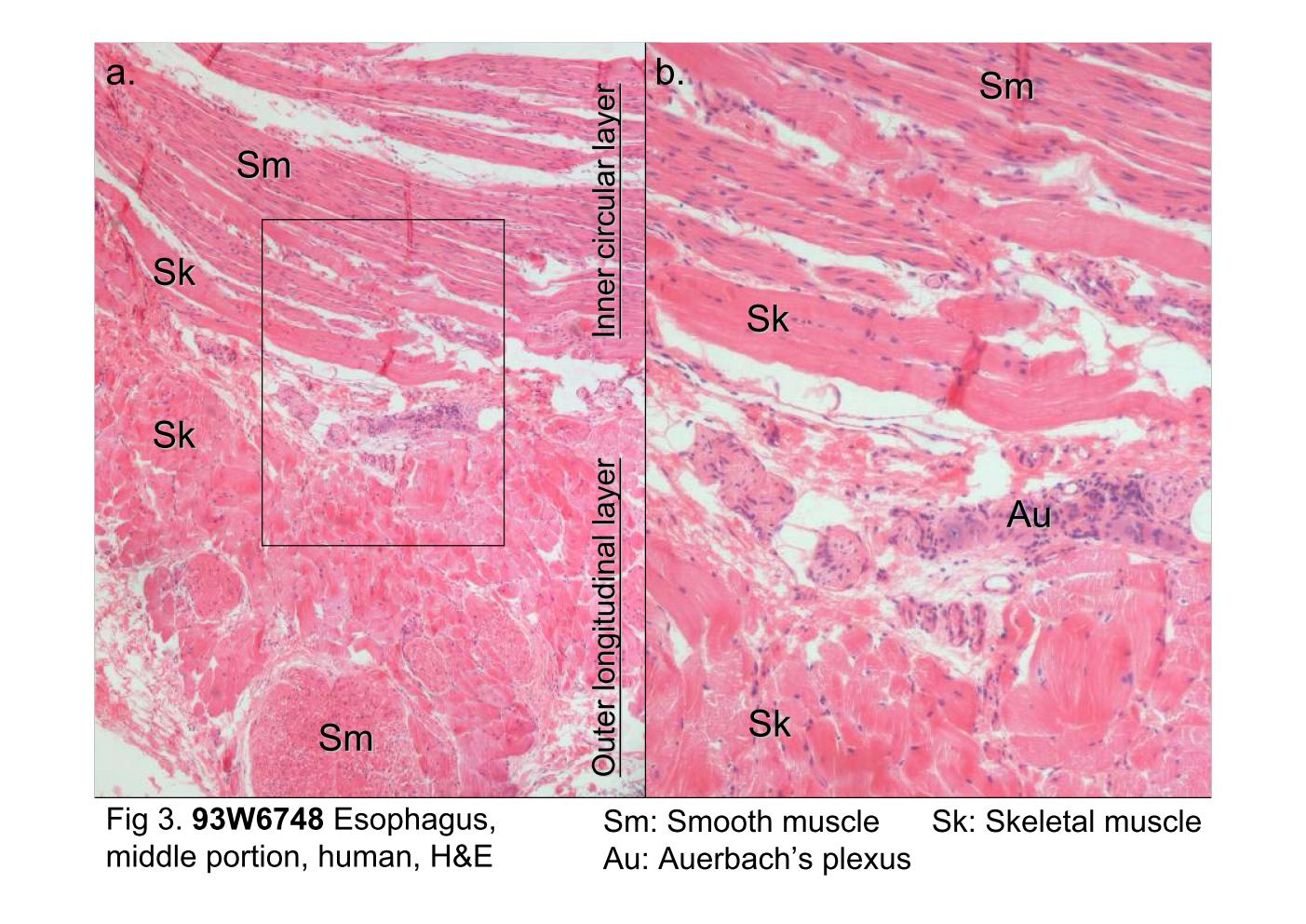
Fig 3. The microscopic structure of the muscularis externa of the esophagus. The muscularis externa consists of two muscle layers, an inner circular layer and an outer longitudinal layer. The esophagus is divided histologically into three regions on the basis of the type of muscle which muscularis externa contains. At the middle third of the esophagus, skeletal muscle (Sk) and smooth muscle (Sm) bundles are mixed and interwoven in the muscularis externa. Located between the two muscle layers is a thin connective tissue layer. Within this connective tissue lies the Auerbach's plexus (Au) containing nerve cell bodies of postganglionic parasympathetic neurons and neurons of the enteric nervous system, as well as blood vessels and lymphatic vessels. The rectangle of Fig 3a is examined at higher magnification in Fig 3b.
block10-1_02block10-1_03block10-1_04block10-1_05block10-1_07block10-1_09block10-1_11block10-1_13block10-1_15block10-1_17block10-1_19

支援訊息