|
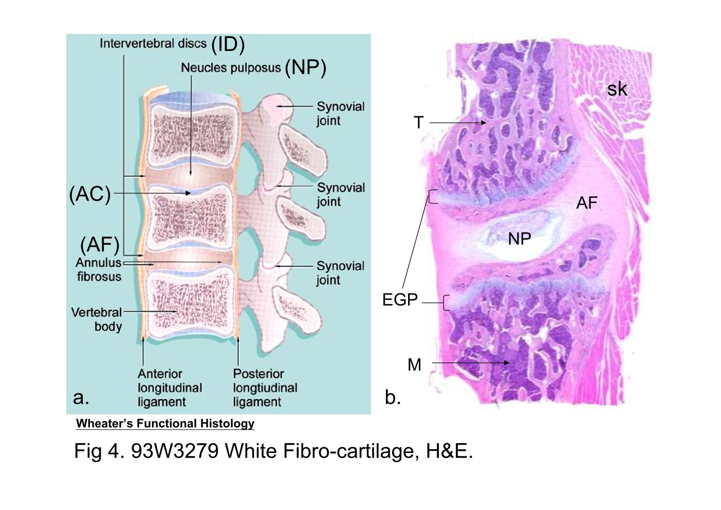 |
| Fig 4a. The intervertebral joints
The intervertebral disc (ID) lies between the adjacent vertebral
bodies. The articular surface of vertebral body is covered by
hyaline articular cartilage (AC). The fibrocartilage of each
intervertebral disc is arranged in concentric rings forming the
annulus fibrosus (AF). Within the disc, there is a central cavity
containing a viscous fluid, the nucleus pulposus (NP). Fig 4b. 93W3279 White Fibro-cartilage (sec.) H&E. This section is prepared from the developing vertebral column. Except the structures mentioned above, the skeletal muscle (sk) and the epiphyseal growth plates (EGP) can be identified. In this low power photomicrograph, the vertebral bodies have been partially replaced by bone trabeculae (T), and the bone cavity is filled with bone marrow (M). |
|||||||||||
支援訊息