Up Block6-1 Prev Next

 上一張相片  下一張相片  索引頁
block6-1_07.jpg - Fig 2. 93W3265 Hyaline Cartilage, H&E.This figure shows a higher-magnification micrograph of a hyalinecartilage with its perichondrium (P). The cartilage appears as anavascular expanse of matrix material and a population of cellscalled chondrocytes (Cc). The chondrocytes produce the matrix;the space each chondrocyte occupies is called a lacuna (L).Hyaline cartilage is surrounded by a thin layer of denseconnective tissue, the perichondrium. The perichondrium servesas a source of new chondrocytes during appositional growth ofthe cartilage. Chondrocytes also undergo interstitial growth inlacunae and form isogenous groups (IP). The chondrocytesproduce the cartilage matrix that shows the dark-staining capsuleor territorial matrix (TM) immediately surrounding the lacunae.The interterritorial matrix (IM) is more removed from theimmediate vicinity of chondrocytes and is less intensely stained.

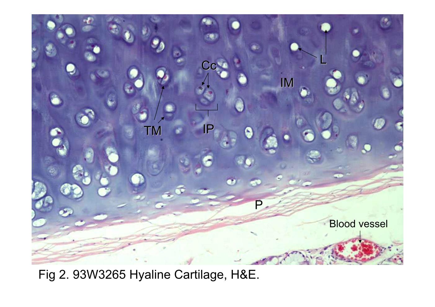
Fig 2. 93W3265 Hyaline Cartilage, H&E. This figure shows a higher-magnification micrograph of a hyaline cartilage with its perichondrium (P). The cartilage appears as an avascular expanse of matrix material and a population of cells called chondrocytes (Cc). The chondrocytes produce the matrix; the space each chondrocyte occupies is called a lacuna (L). Hyaline cartilage is surrounded by a thin layer of dense connective tissue, the perichondrium. The perichondrium serves as a source of new chondrocytes during appositional growth of the cartilage. Chondrocytes also undergo interstitial growth in lacunae and form isogenous groups (IP). The chondrocytes produce the cartilage matrix that shows the dark-staining capsule or territorial matrix (TM) immediately surrounding the lacunae. The interterritorial matrix (IM) is more removed from the immediate vicinity of chondrocytes and is less intensely stained.
block6-1_01block6-1_02block6-1_03block6-1_04block6-1_05block6-1_07block6-1_09block6-1_11block6-1_13block6-1_15block6-1_17

支援訊息