Up Block4 Prev Next

 上一張相片  下一張相片  索引頁
block9_26.jpg - Fig 12. Lung (NO-6-b)A typical bronchiole is shown here. Blood vessels (BV) are adjacent to the bronchiole. The main features of the bronchiolar wall evident in the figure are bundles of smooth muscle (M) and the lining epithelium. The connective tissue is minimal and, at this low magnification, not conspicuous. Nevertheless, the connective tissue is present and separates the muscle into bundles. The connective tissue contains collagenous fibers (CF). Glands are not present in the wall of the bronchiole. Surrounding the bronchiole, are the air spaces or alveoli (A) of the lung.

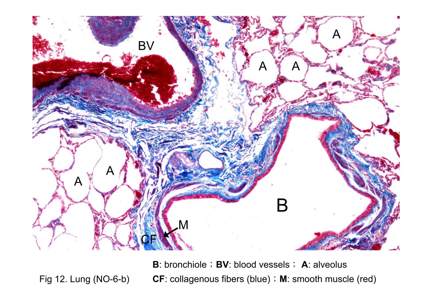
Fig 12. Lung (NO-6-b) A typical bronchiole is shown here. Blood vessels (BV) are adjacent to the bronchiole. The main features of the bronchiolar wall evident in the figure are bundles of smooth muscle (M) and the lining epithelium. The connective tissue is minimal and, at this low magnification, not conspicuous. Nevertheless, the connective tissue is present and separates the muscle into bundles. The connective tissue contains collagenous fibers (CF). Glands are not present in the wall of the bronchiole. Surrounding the bronchiole, are the air spaces or alveoli (A) of the lung.
block9_17block9_19block9_20block9_22block9_24block9_26block9_28block9_30block9_32  

支援訊息