Up Block4 Prev Next

 上一張相片  下一張相片  索引頁
block9_20.jpg - Fig 8 & 9. Trachea (NO-5-a)The wall of the trachea shows the pseudostratified columnar ciliated epithelium (PCE) located on a well-developed basement membrane (BM). A thin lamina propria (LP) and a dense thick submucosa (SM) underlie the respiratory epithelium. Numerous goblet cells (GC) are evident as clear ovoid spaces in the respiratory epithelium. Seromucous glands (tracheal glands) (TG) are seen in the submucosa.

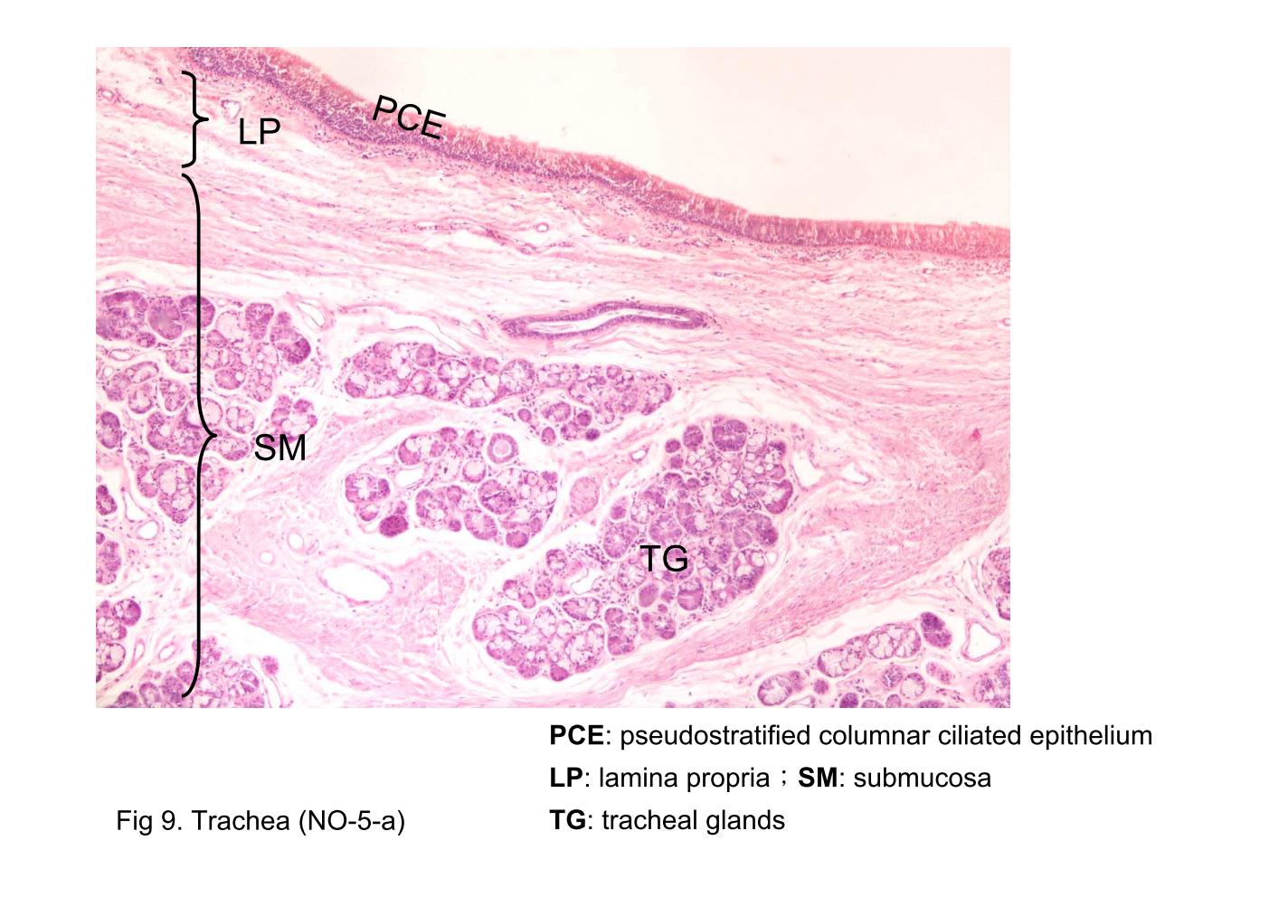
Fig 8 & 9. Trachea (NO-5-a) The wall of the trachea shows the pseudostratified columnar ciliated epithelium (PCE) located on a well-developed basement membrane (BM). A thin lamina propria (LP) and a dense thick submucosa (SM) underlie the respiratory epithelium. Numerous goblet cells (GC) are evident as clear ovoid spaces in the respiratory epithelium. Seromucous glands (tracheal glands) (TG) are seen in the submucosa.
block9_11block9_13block9_15block9_17block9_19block9_20block9_22block9_24block9_26block9_28block9_30

支援訊息